Acridine Orange hydrochloride;(Synonyms: 吖啶橙) 纯度: 99.72%
Acridine Orange hydrochloride 是与核酸结合的细胞渗透性荧光染料,导致光谱发射改变。

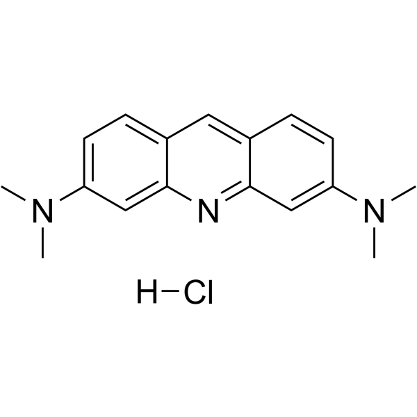
Acridine Orange hydrochloride Chemical Structure
CAS No. : 65-61-2
| 规格 | 价格 | 是否有货 | 数量 |
|---|---|---|---|
| 10;mM;*;1 mL in DMSO | ¥550 | In-stock | |
| 100 mg | ¥500 | In-stock | |
| 200 mg | ; | 询价 | ; |
| 500 mg | ; | 询价 | ; |
* Please select Quantity before adding items.
Acridine Orange hydrochloride 相关产品
bull;相关化合物库:
- Bioactive Compound Library Plus
- Anti-Blood Cancer Compound Library
| 生物活性 |
Acridine Orange hydrochloride is a cell-permeable fluorescent dye that binds to nucleic acids, resulting in an altered spectral emission. |
||||||||||||||||
|---|---|---|---|---|---|---|---|---|---|---|---|---|---|---|---|---|---|
| 体外研究 (In Vitro) |
Acridine Orange has been employed extensively as a cytochemical stain and has shown to stain differentially DNA and RNA, and double-restranded nucleic acids in situ. Acridine Orange can either intercalate into double helical nucleic acids (green fluorescence at 530 nm), or bind electrostatically to phosphate groups of single-stranded molecules (red fluorescence at 640 nm). This unique characteristic makes acridine orange useful for cell-cycle studies[1]. Acridine Orange staining of unfixed cells may be used as a simple, fast means of obtaining information on cell ploidy levels and cell cycle status from DNA measurements (green fluorescence), and cell transcriptional activity from RNA staining (red fluorescence), in human and murine cells lines, peripheral blood and bone marrow specimens from patients with leukemia and mitogenically (phytohemagglutinin) or antigenically (mixed lymphocyte culture) stimulated human peripheral blood cultures[2]. MCE has not independently confirmed the accuracy of these methods. They are for reference only. |
||||||||||||||||
| 分子量 |
301.81 |
||||||||||||||||
| Formula |
C17H20ClN3 |
||||||||||||||||
| CAS 号 |
65-61-2 |
||||||||||||||||
| 中文名称 |
吖啶橙;碱性橙 14;丫啶橙;3,6-双(二甲氨基)吖啶 |
||||||||||||||||
| 运输条件 |
Room temperature in continental US; may vary elsewhere. |
||||||||||||||||
| 储存方式 |
4deg;C, protect from light, stored under nitrogen *In solvent : -80deg;C, 6 months; -20deg;C, 1 month (protect from light, stored under nitrogen) |
||||||||||||||||
| 溶解性数据 |
In Vitro:;
H2O : 50 mg/mL (165.67 mM; ultrasonic and heat to 60°C) DMSO : 25 mg/mL (82.83 mM; Need ultrasonic) 配制储备液
*
请根据产品在不同溶剂中的溶解度选择合适的溶剂配制储备液;一旦配成溶液,请分装保存,避免反复冻融造成的产品失效。 In Vivo:
请根据您的实验动物和给药方式选择适当的溶解方案。以下溶解方案都请先按照 In Vitro 方式配制澄清的储备液,再依次添加助溶剂: ——为保证实验结果的可靠性,澄清的储备液可以根据储存条件,适当保存;体内实验的工作液,建议您现用现配,当天使用; 以下溶剂前显示的百
|
||||||||||||||||
| 参考文献 |
|
| Cell Assay |
Two-step, pH 3.0: Aliquots (0.2 mL, containing approximately 2-5×10⁵ cells) are withdrawn from cultures and are added to 0.5 mL of a solution containing: 0.1% (v/v) Triton X-100, 0.2 M sucrose, 10-4 M EDTA and 2×10⁻² M citrate-phosphate buffer, at pH 3.0. Triton X-100 is included in the various procedures at the indicated pH to increase cell permeability yet maintain cellular integrity. The chelating agent EDTA is used to facilitate RNA denaturation. The cells are stained one minute later by addition of 1 mL of a solution containing 0.002% (20 μg/mL) AO, 0.1 M NaCl and 10-2 M citrate-phosphate buffer, pH 3.8. Cations are included in the staining mixture to ensure staining specificity. The final AO concentration is approximately 4×10⁻⁵ M[2]. MCE has not independently confirmed the accuracy of these methods. They are for reference only. |
|---|---|
| 参考文献 |
|


