H2DCFDA(Synonyms: DCFH-DA 2′,7′-Dichlorodihydrofluorescein diacetate)

H2DCFDA;(Synonyms: DCFH-DA; 2′,7′-Dichlorodihydrofluorescein diacetate) 纯度: 99.81%

H2DCFDA (DCFH-DA) 是一种可渗透细胞的,用于检测细胞内活性氧 (ROS) 的探针 (Ex/Em=488/525 nm)。
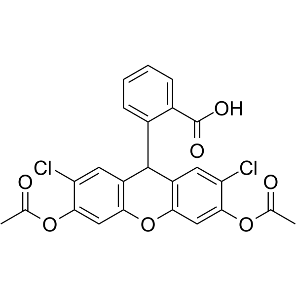
H2DCFDAamp;;(Synonyms: DCFH-DA;  2

H2DCFDA Chemical Structure

CAS No. : 4091-99-0

规格 价格 是否有货 数量
10;mM;*;1 mL in DMSO ¥550 In-stock
50 mg ¥500 In-stock
100 mg ¥700 In-stock
200 mg ; 询价 ;
500 mg ; 询价 ;

* Please select Quantity before adding items.

H2DCFDA 相关产品

bull;相关化合物库:

  • Bioactive Compound Library Plus
  • Immunology/Inflammation Compound Library
  • Metabolism/Protease Compound Library
  • NF-kappa;B Signaling Compound Library
  • Stem Cell Signaling Compound Library
  • Anti-Aging Compound Library
  • Antioxidants Compound Library
  • Oxygen Sensing Compound Library
  • Ferroptosis Compound Library
  • Pyroptosis Compound Library
  • Mitochondria-Targeted Compound Library

生物活性

H2DCFDA (DCFH-DA) is a cell-permeable probe used to detect intracellular reactive oxygen species (ROS) (Ex/Em=488/525 nm)[1].

体外研究
(In Vitro)

Guidelines (Following is our recommended protocol. This protocol only provides a guideline, and should be modified according to your specific needs).
1. H2DCFDA is dissolved in DMSO to obtain a 10 mM stock solutions and further diluted before use.
2. Cells are incubated with 5 µM H2DCFDA solution in PBS in the dark for 30 min at 37°C, then harvested with 0.05% trypsin-EDTA solution, suspended in a fresh medium, and immediately analyzed with flow cytometer.
3. Along with the H2DCFDA probe, if indicated, use ROS-insensitive modification of the fluorescein dye DCFDA as a positive control. The staining procedure is the same as for the H2DCFDA[1].

MCE has not independently confirmed the accuracy of these methods. They are for reference only.

分子量

487.29

Formula

C24H16Cl2O7

CAS 号

4091-99-0

运输条件

Room temperature in continental US; may vary elsewhere.

储存方式

-20deg;C, protect from light

*In solvent : -80deg;C, 6 months; -20deg;C, 1 month (protect from light)

溶解性数据
In Vitro:;

DMSO : 125 mg/mL (256.52 mM; Need ultrasonic)

Ethanol : 20 mg/mL (41.04 mM; Need ultrasonic)

配制储备液
浓度 溶剂体积 质量 1 mg 5 mg 10 mg
1 mM 2.0522 mL 10.2608 mL 20.5217 mL
5 mM 0.4104 mL 2.0522 mL 4.1043 mL
10 mM 0.2052 mL 1.0261 mL 2.0522 mL

*

请根据产品在不同溶剂中的溶解度选择合适的溶剂配制储备液;一旦配成溶液,请分装保存,避免反复冻融造成的产品失效
储备液的保存方式和期限:-80°C, 6 months; -20°C, 1 month (protect from light)。-80°C 储存时,请在 6 个月内使用,-20°C 储存时,请在 1 个月内使用。

In Vivo:

请根据您的实验动物和给药方式选择适当的溶解方案。以下溶解方案都请先按照 In Vitro 方式配制澄清的储备液,再依次添加助溶剂:

——为保证实验结果的可靠性,澄清的储备液可以根据储存条件,适当保存;体内实验的工作液,建议您现用现配,当天使用; 以下溶剂前显示的百
分比是指该溶剂在您配制终溶液中的体积占比;如在配制过程中出现沉淀、析出现象,可以通过加热和/或超声的方式助溶

  • 1.

    请依序添加每种溶剂:;10% DMSO ;; 40% PEG300 ;; 5% Tween-80 ;; 45% saline

    Solubility: 2.08 mg/mL (4.27 mM); Suspended solution; Need ultrasonic

    此方案可获得 2.08 mg/mL (4.27 mM) 的均匀悬浊液,悬浊液可用于口服和腹腔注射。

    以 1 mL 工作液为例,取 100 μL 20.8 mg/mL 的澄清 DMSO 储备液加到 400 μL PEG300 中,混合均匀;向上述体系中加入50 μL Tween-80,混合均匀;然后继续加入 450 μL生理盐水定容至 1 mL。

    将 0.9 g 氯化钠,完全溶解于 100 mL ddH₂O 中,得到澄清透明的生理盐水溶液

  • 2.

    请依序添加每种溶剂:;10% DMSO ;; 90% (20% SBE-β-CD in saline)

    Solubility: ≥ 2.08 mg/mL (4.27 mM); Clear solution

    此方案可获得 ≥ 2.08 mg/mL (4.27 mM,饱和度未知) 的澄清溶液。

    以 1 mL 工作液为例,取 100 μL 20.8 mg/mL 的澄清 DMSO 储备液加到 900 μL 20% 的 SBE-β-CD 生理盐水水溶液中,混合均匀。

    将 2 g 磺丁基醚 β-环糊精加入 5 mL 生理盐水中,再用生理盐水定容至 10 mL,完全溶解,澄清透明
  • 3.

    请依序添加每种溶剂:;10% DMSO ;; 90% corn oil

    Solubility: ≥ 2.08 mg/mL (4.27 mM); Clear solution

    此方案可获得 ≥ 2.08 mg/mL (4.27 mM,饱和度未知) 的澄清溶液,此方案不适用于实验周期在半个月以上的实验。

    以 1 mL 工作液为例,取 100 μL 20.8 mg/mL 的澄清 DMSO 储备液加到 900 μL玉米油中,混合均匀。

*以上所有助溶剂都可在 MCE 网站选购。
参考文献
  • [1]. Lyublinskaya OG, et al. Redox environment in stem and differentiated cells: A quantitative approach. Redox Biol. 2017 Aug;12:758-769.

Kinase Assay

ROS Measurements[1]
For the detection of intracellular ROS level ,ROS-sensitive probe H2DCFDA is used. Adherent cells (ESCs, difESCs, eMSCs, HeLa, U118) are incubated with 5 µM staining solution in PBS in the dark for 30 min at 37°C, then harvested with 0.05% trypsin-EDTA solution, suspended in a fresh medium, and immediately analyzed with flow cytometer. Lymphocytes, both control and PHA-activated, are resuspended in PBS, incubated with 5 µM of H2DCFDA in the dark for 30 min at 37°C, and immediately analyzed. Along with the H2DCFDA probe, if indicated, ROS-insensitive modification of the fluorescent dye DCFDA is used. The staining procedure is the same as for the H2DCFDA[1].

MCE has not independently confirmed the accuracy of these methods. They are for reference only.

参考文献
  • [1]. Lyublinskaya OG, et al. Redox environment in stem and differentiated cells: A quantitative approach. Redox Biol. 2017 Aug;12:758-769.