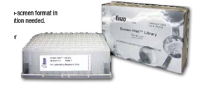
SCREEN-WELL® Autophagy library Screen-Well 自噬 化合物库 品牌:Enzo


SCREEN-WELL® Autophagy library

Screen-Well 自噬 化合物库

品牌:Enzo
CAS No.:
储存条件:-80℃
纯度:
产品编号

(生产商编号)

等级 规格 运输包装 零售价(RMB) 库存情况 参考值

BML-2837-0500

1 Library 咨询


* 干冰运输、大包装及大批量的产品需酌情添加运输费用


* 零售价、促销产品折扣、运输费用、库存情况、产品及包装规格可能因各种原因有所变动,恕不另行通知,确切详情请联系上海金畔生物科技有限公司。

日本同仁化学DAPRed – Autophagy Detection 细胞自噬检测试剂货号:D677| DOJINDO

上海金畔生物科技有限公司代理日本同仁化学 DOJINDO代理商全线产品,欢迎访问官网了解更多信息

DAPRed – Autophagy Detection 细胞自噬检测试剂货号:D677
细胞自噬检测试剂盒
DAPRed – Autophagy Detection
商品信息
储存条件:
0-5度保存,避光
运输条件:
室温
特点:

● 操作流程简便

● 与LC3结果高度一致

● 可以动态观察细胞自噬

下载说明书
产品文献
SDS下载

选择规格:
5nmol

现货

操作简便
活细胞检测
自噬小体检测
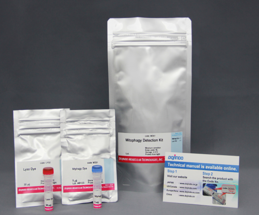
线粒体自噬检测(点击查看)

活动进行中产品概述原理试剂概要操作简便实验例DAPRed荧光光谱常见问题Q&A参考文献
活动进行中

订购满5000元,200元礼品等你拿

凑单关联产品TOP5

NO.1. DALGreen – Autophagy Detection 细胞自噬检测

NO.2. Cell Counting Kit-8 细胞增殖毒性检测

NO.3. Caspase-3 Assay Kit-Colorimetric- 细胞凋亡检测

NO.4. Liperfluo 细胞脂质过氧化物检测

NO.5. Mitophagy Detection Kit 线粒体自噬检测

产品概述

细胞自噬是细胞内损坏的蛋白质或细胞器降解和循环利用的过程。
DAPRed是一种小分子荧光染料,可以用来检测自噬体及自噬溶酶体。由于其特殊的结构,在自噬体形成双层膜结构时染料可以进入其中,并在疏水环境中产生荧光。
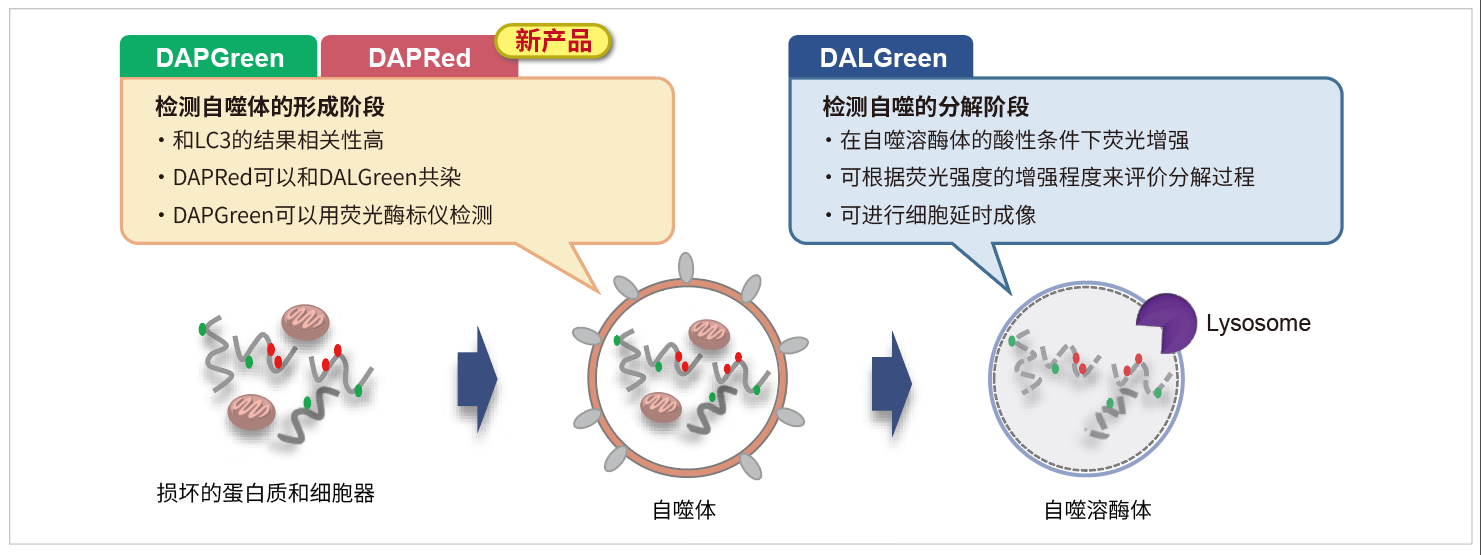
DAPRed可以进入自噬体膜而发出荧光;而DALGreen可以在自噬溶酶体产生阶段发出荧光。因此DAPRed,DALGreen可以检测从自噬体的形成到自噬溶酶体的融合以及内容物分解的整个过程。

原理

1622787436478709.png

当形成自噬体膜时,DAPRed可以掺入其中,并在脂溶性环境中产生荧光。

DAPRed的检测结果与细胞自噬标志物LC3的结果有很高的相关性。

1622787557957820.png

试剂概要

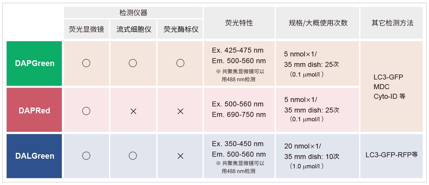
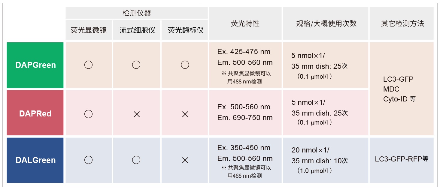
使用前,请确认所用仪器可以检测到的荧光特性。

1622787655866132.png

操作简便

操作流程只有一步-加入试剂
无需基因转染。只需向准备好的细胞加入DAPRed染料,即可方便快捷的进行荧光检测。

1612415620431352.jpg

实验例

DAPRed和DAL Green的共染
用自噬体荧光试剂DAPRed和自噬溶酶体荧光试剂DALGreen对HeLa细胞进行共染后,通过饥饿培养诱导自噬。

1622788180693956.png

·结果

在不含氨基酸的培养基中培养的HeLa细胞,DAPRed和DALGreen荧光增强。

·检测条件
DAPRed:EX. 561 nm / Em. 600-700 nm
DALGreen:EX. 488 nm / Em. 500-563 nm
比例尺 :20 μm

·自噬诱导条件
用DAPRed和DALGreen染色后的HeLa细胞分别在增殖型培养基和不含氨基酸的培养基中培养5 h后,用共聚焦显微镜观察。

DAPRed荧光光谱

1607288752418470.png

常见问题Q&A

Q1: DAPRed Working Solution的稳定性如何?

A1: 无法长期保存,需要现配现用
Q2: DAPRed DMSO Stocking Solution的稳定性如何?

A2: 配制后请于-20℃保存,一个月内可保持稳定。另外建议根据用量分装保存。
Q3: 推荐使用的滤光片?

A3: 推荐的滤光片如下:
激发波长:500-560 nm

发射波长:690-750 nm

Q4: 如何确定细胞自噬探针DAPRed的最佳浓度?

A4:由于本试剂的特性,如果试剂的浓度太高或太低都会导致诱导自噬的样品组与未诱导自噬的对照组之间的差别不明显。建议参考以下信息摸索试剂的最佳浓度:

探针的最佳浓度根据细胞的种类而不尽相同。以DAPRed为例可以考虑从最低浓度(可以以0.05μmol/l作为参考)开始分别多个梯度至摸索至最高浓度(可以以0.4 μmol/l作为参考)的步骤进行摸索。

1622108968137374.png

参考例

我们公司对HeLa, HepG2, CHO细胞的最佳浓度进行了摸索。DAPRed以下列浓度进行染色,并在无氨基酸的培养基中培养以诱导自噬。下表中红色字体的浓度可明显观察到实验组与空白组的差异。

[HeLa细胞]

1612416674568578.jpg

<检测条件>放大倍率:20倍; 激发波长:Ex:561 nm;发射波长:Em:600-700 nm

[HepG2细胞]

1612416807368714.jpg

<检测条件>放大倍率:20倍; 激发波长:Ex:561 nm;发射波长:Em:600-700 nm

[CHO细胞]

1612416864229024.jpg

<检测条件>放大倍率:20倍; 激发波长:Ex:561 nm;发射波长:Em:600-700 nm

Q5: 自噬有哪些途径?DAPRed可以检测到哪些状态?

A5: 众所周知,自噬根据其分子机制可以分为两种:一种是依赖于ATG5的传统自噬(LC3发生变化),另一种则是非依赖于ATG的选择性自噬(LC3形式的转化并未发生)。
当形成自噬体膜时,DAPRed可以掺入其中,并在脂溶性环境中产生荧光。因此DAPRed可以检测自噬体的状态。

*参考资料:发现新的自噬机制Shigeomi Shimizu

https://www.dojindo.co.jp/letterj/160/review/01.html

文献链接:http://dx.doi.org/10.14348/molcells.2018.2215

▶对于首次检测的细胞类型和实验条件,请参考FAQ[如何确定细胞自噬探针DAPRed的最佳浓度]。

参考文献

No.

检测样品

检测仪器

引用(含链接)

1)

细胞
(HUVEC)

荧光显微镜

X. Chen, X. Yan, J. Liu and L. Zhang, ” Chaiqi decoction ameliorates vascular endothelial injury in metabolic syndrome by upregulating autophagy.”, Am. J. Transl. Res., 2020,12(9), 4902.

2)

细胞
(HeLa)

荧光显微镜

H. Fang , S. Geng, M. Hao, Q. Chen, M. Liu, C. Liu, Z. Tian, C. Wang, T. Takebe, J-L Guan, Y. Chen, Z. Guo, W. He and J. Diao, “Simultaneous Zn2+ tracking in multiple organelles using super-resolution morphology-correlated organelle identification in living cells “, Nat Commun, 2021, 12(1), 109. 10.1016/j.envpol.2019.07.105.

3)

细胞
(HCT116; HCT8)

荧光显微镜

H. Sun, R. Wang, Y. Liu, H. Mei and X. Liu , “USP11 induce resistance to 5-Fluorouracil in Colorectal Cancer through activating autophagy by stabilizing VCP”, J Cancer , 2021, 12(8), 2317.

4)

细胞
(MEF)

荧光显微镜

M. Yagi, T. Toshima, R. Amamoto, Y. Do, H. Hirai, D. Setoyama, D. Kang and T. Uchiumi, “Mitochondrial translation deficiency impairs NAD+-mediated lysosomal acidification”, EMBO J, 2021, doi:10.15252/embj.2020105268.

关联产品

线粒体自噬—Mitophagy Detection Kit
线粒体自噬检测试剂盒

DALGreen – Autophagy Detection 细胞自噬检测试剂
DALGreen – 细胞自噬荧光探针

DAPGreen – Autophagy Detection 细胞自噬检测试剂
DAPGreen – 细胞自噬荧光探针

Mtphagy Dye试剂
Mtphagy Dye

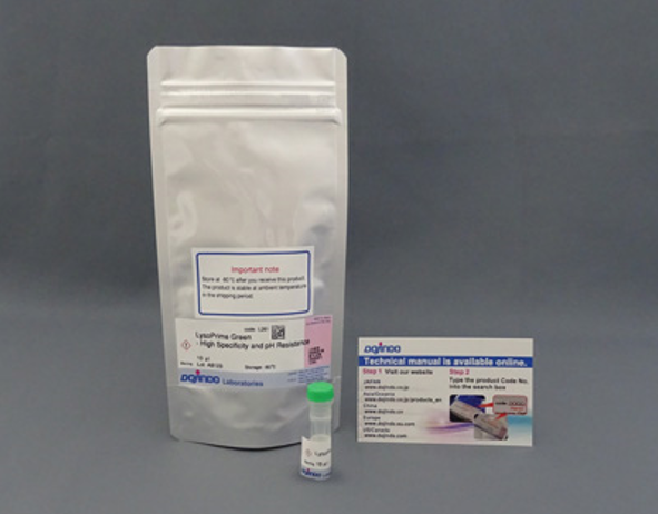
LysoPrime Green – High Specificity and pH Resistance
溶酶体染色试剂Green
在线小工具
实验工具|稀释计算器|摩尔浓度计算器

日本同仁化学DAPGreen – Autophagy Detection 细胞自噬检测试剂货号:D676| DOJINDO

上海金畔生物科技有限公司代理日本同仁化学 DOJINDO代理商全线产品,欢迎访问官网了解更多信息

DAPGreen – Autophagy Detection 细胞自噬检测试剂货号:D676
DAPGreen – 细胞自噬荧光探针
DAPGreen – Autophagy Detection
商品信息
储存条件:-20度保存,避光
运输条件:室温

特点:

● 操作流程简便

● 与LC3结果高度一致

● 可以动态观察细胞自噬

下载说明书
产品文献
SDS下载

选择规格:
5nmol

现货

操作简便

荧光/流式/荧光酶标仪可测

自噬小体检测

线粒体自噬检测(点击查看)

DAPGreen &#8211; Autophagy Detection 细胞自噬检测试剂货号:D676

DAPGreen &#8211; Autophagy Detection 细胞自噬检测试剂货号:D676

活动进行中
产品概述
原理
试剂概要
操作简便
实验例
DAPGreen荧光光谱
常见问题Q&A
参考文献

活动进行中

订购满5000元,200元礼品等你拿

凑单关联产品TOP5

NO.1.    Cell Counting Kit-8    细胞增殖毒性检测

NO.2.    Mitophagy Detection Kit    线粒体自噬检测

NO.3.    Calcein-AM/PI Double Staining Kit    活死细胞双染

NO.4.    Liperfluo    细胞脂质过氧化物检测

NO.5.    Annexin V, FITC Apoptosis Detection Kit    细胞凋亡检测

产品概述

DAPGreen是一种小分子荧光染料,可以用来检测自噬体及自噬溶酶体。由于其特殊的结构,在自噬体形成双
层膜结构时染料可以进入其中,并在疏水环境中产生荧光。DAPGreen具有很好的细胞透膜性,通过荧光显微
镜可进行活细胞荧光成像,也可使用流式细胞仪进行定量检测。

原理

DAPGreen &#8211; Autophagy Detection 细胞自噬检测试剂货号:D676

当形成自噬体膜时,DAPGreen可以掺入其中,并在脂溶性环境中产生荧光。

DAPGreen的检测结果与细胞自噬标志物LC3的结果有很高的相关性。

DAPGreen &#8211; Autophagy Detection 细胞自噬检测试剂货号:D676

试剂概要

DAPGreen不仅可以用荧光显微镜检测,还可以使用流式细胞仪进行检测。

同时也实现了用荧光酶标仪进行检测,您可以根据自己的实验条件,使用不同的仪器进行检测。

DAPGreen &#8211; Autophagy Detection 细胞自噬检测试剂货号:D676                                                                                                                                                                                                        *DAPGreen和DALGreen不能共染

操作简便

操作流程只有一步-加入试剂

只需向准备好的细胞加入DAPGreen染料,即可方便快捷的进行荧光检测。

DAPGreen &#8211; Autophagy Detection 细胞自噬检测试剂货号:D676

实验例

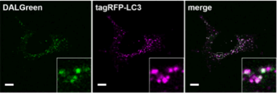
与LC3高度相关           
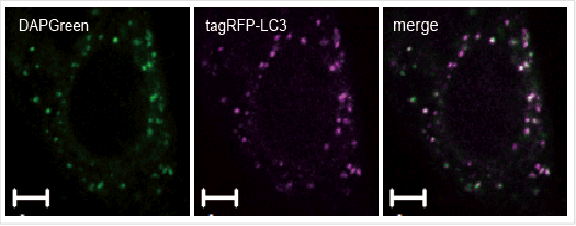
与自噬标准物LC3的细胞共染实验结果的比较。
DAPGreen &#8211; Autophagy Detection 细胞自噬检测试剂货号:D676

检测条件:DAPGreen:Ex. 488 nm / Em. 500-563 nm

比例尺:10 μm
将DAPGreen加入已表达tagRFP-LC3的Hela细胞中,用雷帕霉素(Rapamycin)诱导自噬4 h后,用共聚焦显微镜观察DAPGreen和RFP的荧光成像。结果DAPGreen与tagRFP-LC3的染色部位高度一致。

与Lamp-1共染                                                               DAPGreen &#8211; Autophagy Detection 细胞自噬检测试剂货号:D676
对已经表达Lamp1-tagRFP的MEF细胞进行DAPGreen共染实验。结果显示,DAPGreen的染色部位与溶酶体膜蛋白标记物Lamp 1的位置高度一致。(比例尺:10 μm)

实验的详细情况请参考如下论文:
“Small fluorescent molecules for monitoring autophagic flux”, FEBS Letters., 2018, 592, (4), 559–567.

流式细胞仪的定量分析

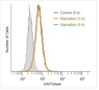
自噬诱导后,通过流式细胞仪检测DAPGreen的荧光。

DAPGreen &#8211; Autophagy Detection 细胞自噬检测试剂货号:D676

检测条件:
检测波长:Ex. 488 nm / Em. 500-560 nm

DAPGreen染色HeLa细胞后,在不含氨基酸的培养基中培养0、3和6 h,用流式细胞仪检测。结果,在饥饿诱导3 h后可以检测到更强的荧光信号。

荧光酶标仪的定量检测
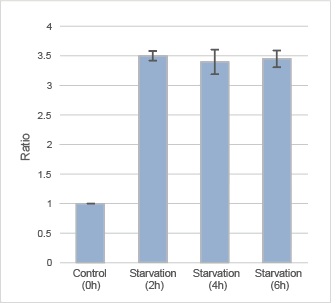
用荧光酶标仪检测自噬诱导后DAPGreen的荧光。
DAPGreen &#8211; Autophagy Detection 细胞自噬检测试剂货号:D676

检测条件

检测波长:Ex. 450 nm / Em. 535 nm

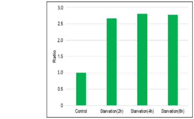
DAPGreen染色HeLa细胞后,在不含氨基酸的培养基中分别培养0、2、4、6 h,用荧光酶标仪检测。结果饥饿诱导2 h检测荧光,饥饿诱导组的荧光强度大约是对照组的3.5倍。

有关检测操作的详细信息,请参考FAQ“使用荧光酶标仪进行定量分析的检测条件是什么?”。

应用例:荧光显微镜的数值化

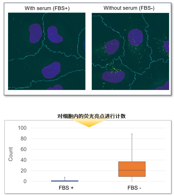
96孔板中接种HeLa细胞,并用DAPGreen染色后,分别更换含有血清和不含血清的培养基后继续培养。培养后分别通过视野内的平均荧光强度进行计算。结果发现,不含血清的培养基培养的细胞中DAPGreen的荧光强度更高。

DAPGreen &#8211; Autophagy Detection 细胞自噬检测试剂货号:D676

<检测条件>

Ex:488 nm, Em: 500-550 nm

物镜: CFI Plan Apochromat VC 20x

拍摄模式: Resonant Scanner

XY分辨率:512×512

<使用装置>

荧光显微镜:Nikon 激光共聚焦显微镜A1R

分析软件:NIS-Elements

<实验步骤>

1. 将HeLa细胞播种于96孔板中并培养。

2. 去除培养基,用无血清培养基清洗1次。

3. 添加配置好的DAPGreen working solution ,37℃ 培养30 min。

4. 去除培养基,用无血清培养基清洗2次。

5. 诱导自噬的孔中加入无血清培养基,正常孔中加入正常培养基。37℃培养6小时(之后用4%PFA固定)。

6. 荧光显微镜观察。

按上述条件拍摄后,再用100倍物镜放大后拍摄,对细胞内的DAPGreen的荧光信号进行统计。结果显示,含有血清的培养基培养的细胞中平均每个细胞有1.5个信号,而不含血清的培养基培养的细胞中平均每个细胞有27个信号。

DAPGreen &#8211; Autophagy Detection 细胞自噬检测试剂货号:D676

<检测条件>

Ex : 488 nm, Em : 500-550 nm

物镜: CFI Plan Apochromat TIRF 100xC Oil

拍摄模式: Galvano Scanner

XY分辨率:512×512

<使用装置>

荧光显微镜:Nikon 激光共聚焦显微镜A1R

分析软件:NIS-Elements

*本数据由DAPGreen的使用客户友情提供。

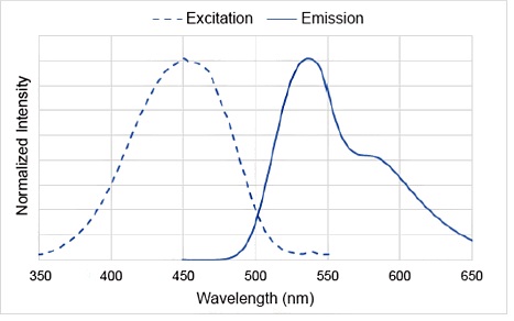
DAPGreen荧光光谱

DAPGreen &#8211; Autophagy Detection 细胞自噬检测试剂货号:D676

激发滤光片:425-475 nm
荧光滤光片:500-560 nm

常见问题Q&A

Q1: DAP Green working solution的稳定性如何?
A1:无法长期保存,需要现配现用
Q2: DMSO stocking solution 的稳定性如何?
A2:配制后请于-20℃保存,一个月内可保持稳定。另外建议根据用量分装保存。
Q3: 推荐使用的滤光片?
A3:激发波长:425-475 nm

发射波长:500-560 nm

利用激光共聚焦显微镜的488 nm激发波长也可以检测,请参考我们公司网站产品页面的实验例。

Q4: 在进行延时成像时有什么要注意的地方吗?
4:为了确定最佳实验条件,请先进行预实验。

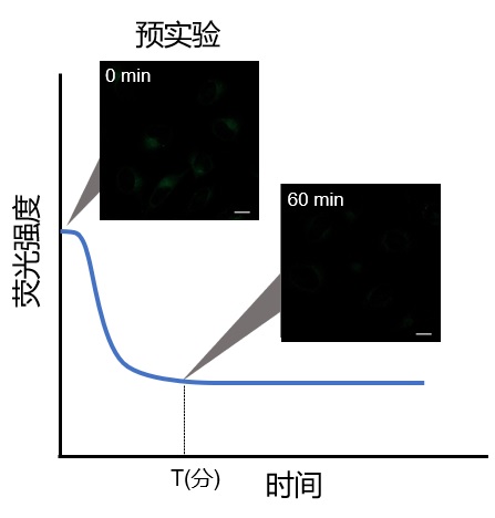
由于试剂的特性,刚刚染色后有荧光值升高的趋势,因此请参照以下步骤进行预实验和延时成像。

DAPGreen &#8211; Autophagy Detection 细胞自噬检测试剂货号:D676

1. 预实验

使用对照细胞(不诱导自噬的细胞)。

根据说明书的步骤用 Working Soluiton染色后,用培养基洗涤2次。

加入正常培养基后,观察荧光随时间的变化。

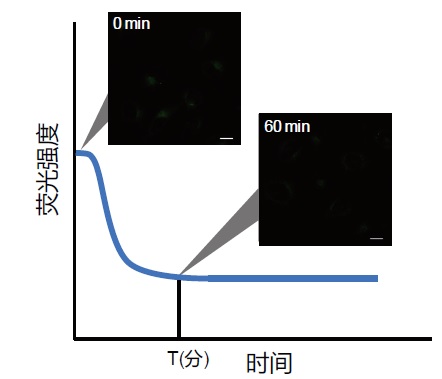
如下图所示,染色后的细胞在荧光强度阶段性降低之后,确认荧光强度变化趋于稳定的时间段(图中的T)。

※条件可能因细胞种类而异。

(参考)

HeLa细胞染色约60分钟后,荧光会趋于稳定(DAPGreen)。

DAPGreen &#8211; Autophagy Detection 细胞自噬检测试剂货号:D676

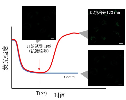
2. 延时染色成像

– 细胞用Working Solution染色后,在培养基中37℃培养。

※培养时间为预实验中摸索出的染色时间。验中摸索出的染色时间。

※染色后不要立刻进行自噬诱导。

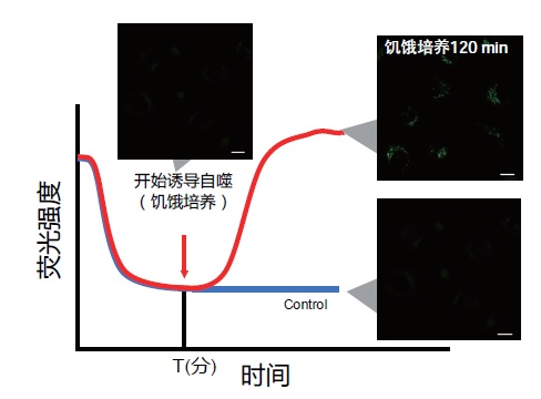
-培养后进行自噬诱导并开始延时染色成像。

(参考)

用DAPGreen对HeLa细胞进行染色,在正常的培养基中培养60分钟(预实验中摸索的时间)后,进行自噬诱导。

Q5: 如何确定细胞自噬探针DAPGreen的最佳浓度?
A5: 由于本试剂的特性,如果试剂的浓度太高或太低都会导致诱导自噬的样品组与未诱导自噬的对照组之间的差别不明显。建议参考以下信息摸索试剂的最佳浓度:

探针的最佳浓度根据细胞的种类而不尽相同。以DAPGreen为例可以考虑从最低浓度(可以以0.05 μmol/l作为参考)开始分别多个梯度至摸索至最高浓度(可以以0.4 μmol/l作为参考)的步骤进行摸索。

参考例:

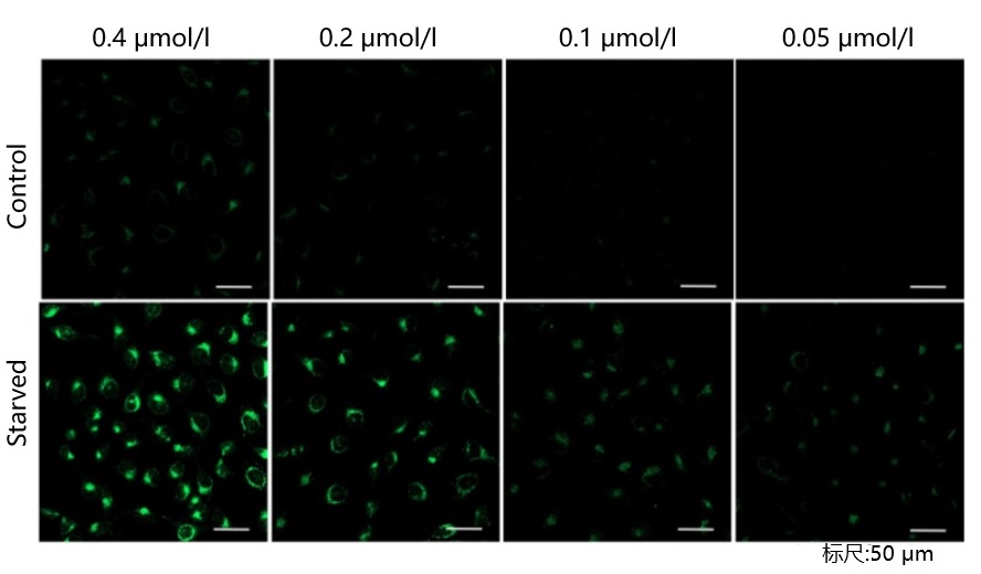
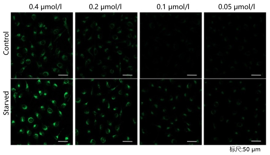
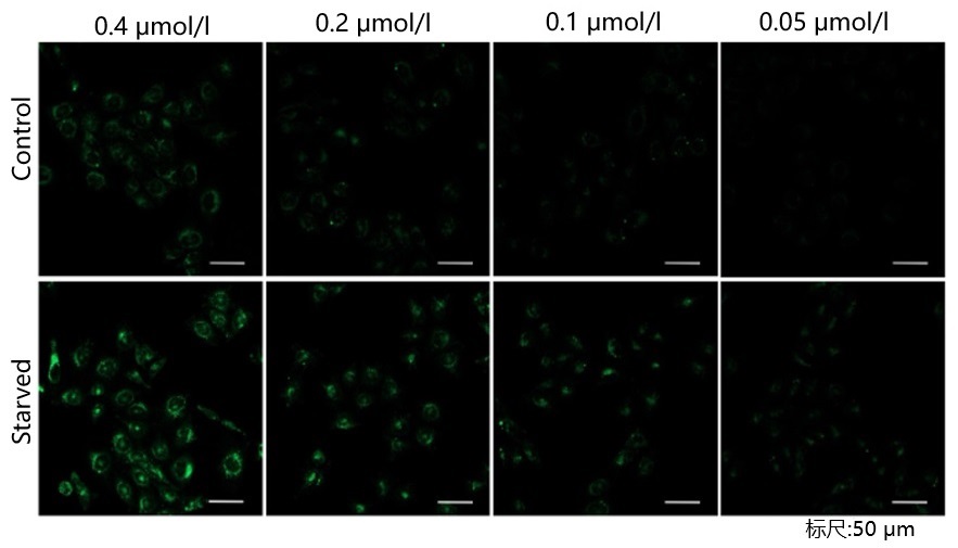
我们公司对HeLa, HepG2, CHO细胞的最佳浓度进行了摸索。DAPGreen以下列浓度进行染色,并在无氨基酸的培养基中培养以诱导自噬。下表中红色字体的浓度可明显观察到实验组与空白组的差异。

[HeLa细胞]

DAPGreen &#8211; Autophagy Detection 细胞自噬检测试剂货号:D676

<检测条件>放大倍率:20倍; 激发波长:Ex:488 nm;发射波长:Em:500-563 nm

[HepG2细胞]

DAPGreen &#8211; Autophagy Detection 细胞自噬检测试剂货号:D676

<检测条件>放大倍率:20倍; 激发波长:Ex:488 nm;发射波长:Em:500-563 nm

[CHO细胞]

DAPGreen &#8211; Autophagy Detection 细胞自噬检测试剂货号:D676

<检测条件>放大倍率:20倍; 激发波长:Ex:488 nm;发射波长:Em:500-563 nm

细胞种类 DAPGreen浓度
Hela 0.4 μmol/l 0.2 μmol/l 0.1 μmol/l 0.05 μmol/l
HepG2 0.4 μmol/l 0.2 μmol/l 0.1 μmol/l 0.05 μmol/l
CHO 0.4 μmol/l 0.2 μmol/l 0.1 μmol/l 0.05 μmol/l
Q6: 使用荧光酶标仪进行定量分析的检测条件是什么?
A6:以下是使用HeLa细胞进行检测的实验例。

将DAPGreen染色的HeLa细胞在不含氨基酸的培养基中分别培养0、2、4、6 h,用荧光酶标仪检测。

<操作>

1)将细胞接种在透明底的黑板上(HeLa细胞,1.6×104 cells/孔,100 µl/孔)

2)在37°C,5% CO2培养箱过夜培养

3)去除上清液后,在培养基中加入100 µl配制好的Working Solution(DAPGreen:0.1 µmo/l)。

4)在37°C,5% CO2培养箱培养30 min

5)用100 µl培养基清洗细胞2次

6)加入100 µl饥饿培养基(Waco,Code:048-33575)

7)在37°C下培养各时间点

8)用荧光酶标仪(TECAN,Infinite Pro M200)检测(Ex/Em = 450 nm/530 nm)

(0 h对照组用HBSS代替检测)

<检测条件>波长:Ex. 450 nm/Em. 535 nm

饥饿诱导2 h检测荧光,确认到饥饿诱导组的荧光强度大约是对照组的2.5倍。DAPGreen &#8211; Autophagy Detection 细胞自噬检测试剂货号:D676

Q7: 自噬有哪些途径?DAPGreen可以检测到哪些状态?
A7:众所周知,自噬根据其分子机制可以分为两种:一种是依赖于ATG5的传统自噬(LC3发生变化),另一种则是非依赖于ATG的选择性自噬(LC3形式的转化并未发生)。

当形成自噬体膜时,DAPGreen可以掺入其中,并在脂溶性环境中产生荧光。因此DAPGreen可以检测自噬体的状态。

*参考资料:发现新的自噬机制Shigeomi Shimizu

https://www.dojindo.co.jp/letterj/160/review/01.html

文献链接:http://dx.doi.org/10.14348/molcells.2018.2215

▶对于首次检测的细胞类型和实验条件,请参考FAQ[如何确定细胞自噬探针DAPGreen的最佳浓度]。

参考文献

No. 检测样品 检测仪器 引用(含链接)
1) 细胞
(HeLa, MEF)
荧光显微镜 H. Iwashita, H. T. Sakurai, N. Nagahora, M. Ishiyama, K. Shioji, K. Sasamoto, K. Okuma, S. Shimizu, and Y. Ueno, “Small fluorescent molecules for monitoring autophagic flux.”, FEBS Letters., 2018, 592, (4), 559–567.
2) 细胞
(HepG2; Huh-7)
荧光显微镜;流式细胞仪  L. Hu, T. Zhang, D. Liu, G. Guan, J. Huang, P. Proksch, X. Chen and W. Lin, “Notoamide-type alkaloid induced apoptosis and autophagy via a P38/JNK signaling pathway in hepatocellular carcinoma cells”, RSC Adv., 2019, 9, 19855.
3) 细胞
(HepG2)
荧光显微镜 Q. Chu, S. Zhang, M. Chen, W. Han, R. Jia, W. Chen and X. Zheng, “Cherry Anthocyanins Regulate NAFLD by Promoting Autophagy Pathway”, Oxid Med Cell Longev., 2019,DOI:10.1155/2019/4825949.
4) 细胞
(HLMVEC)
荧光显微镜 Q. Chu, S. Zhang, M. Chen, W. Han, R. Jia, W. Chen and X. Zheng, “Cherry Anthocyanins Regulate NAFLD by Promoting Autophagy Pathway”, Oxid Med Cell Longev., 2019,DOI:10.1155/2019/4825949.
5) 细胞
(HeLa)
荧光显微镜 F. Hongbao,Y. Shankun, C. Qixin, L. Chunyan, C. Yuqi, G. Shanshan, B. Yang, T. Zhiqi, L. Z. Amanda, T. Takanori, C.Yuncong, G. Zijian, H. Weijiang and D. Jiajie , “De Novo-Designed Near-Infrared Nanoaggregates for Super-Resolution Monitoring of Lysosomes in Cells, in Whole Organoids, and in Vivo.”, ACS Nano, 2019, 13, (12), 1446.
6) 细胞
(HeLa; A375)
流式细胞仪 B. Yang, L. Ding, Y. Chen and J. Shi, “Augmenting Tumor-Starvation Therapy by Cancer Cell Autophagy Inhibition”, Adv. Sci., 2020,DOI:10.1002/advs.201902847.
7) 细胞
(PC12)
荧光显微镜(超分辨率) Y. Tan, L. Yin, Z. Sun, S. Shao, W. Chen, X. Man,Y. Du and Y. Chen, “Astragalus polysaccharide exerts anti-Parkinson via activating the PI3K/AKT/mTOR pathway to increase cellular autophagy level in vitro.”, Int. J. Biol. Macromol., 2020, DOI:10.1016/j.ijbiomac.2020.02.282.
8) 细胞
(Wild type Hepa 1-6)
荧光显微镜

(用ImageJ软件数值化)

J. Kim, W.Y.Chee, N. Yabuta, K. Kajiwara, S. Nada and M. Okada, “Atg5-mediated autophagy controls apoptosis/anoikis via p53/Rb pathway in naked mole-rat fibroblasts”, Biochem. Biophys. Res. Commun., 2020, 22, DOI:10.1016/j.bbrc.2020.05.083.
9) 细胞
(小鼠皮肤成纤维细胞)
流式细胞仪 J. Kim, W.Y.Chee, N. Yabuta, K. Kajiwara, S. Nada and M. Okada, “Atg5-mediated autophagy controls apoptosis/anoikis via p53/Rb pathway in naked mole-rat fibroblasts”, Biochem. Biophys. Res. Commun., 2020, 22, DOI:10.1016/j.bbrc.2020.05.083.
10) 细胞
(HeLa)
荧光显微镜(超分辨率) Q. Chen, M. Hao, L. Wang, L. Li, Y. Chen, X. Shao, Z. Tian, R. A. Pfuetzner, Q. Zhong, A. T. Brunger, J. Guan and J. Diao, “Prefused lysosomes cluster on autophagosomes regulated by VAMP8”,2021, doi:10.1038/s41419-021-04243-0.

关联产品

DAPGreen &#8211; Autophagy Detection 细胞自噬检测试剂货号:D676
线粒体自噬—Mitophagy Detection Kit
线粒体自噬检测试剂盒

DAPGreen &#8211; Autophagy Detection 细胞自噬检测试剂货号:D676
DALGreen – Autophagy Detection 细胞自噬检测试剂
DALGreen – 细胞自噬荧光探针

DAPGreen &#8211; Autophagy Detection 细胞自噬检测试剂货号:D676
DAPRed – Autophagy Detection 细胞自噬检测试剂
细胞自噬检测试剂盒

DAPGreen &#8211; Autophagy Detection 细胞自噬检测试剂货号:D676
Mtphagy Dye试剂
Mtphagy Dye

DAPGreen &#8211; Autophagy Detection 细胞自噬检测试剂货号:D676
LysoPrime Green – High Specificity and pH Resistance
溶酶体染色试剂Green

在线小工具

实验工具|稀释计算器|摩尔浓度计算器

日本同仁化学DALGreen – Autophagy Detection 细胞自噬检测试剂货号:D675| DOJINDO

上海金畔生物科技有限公司代理日本同仁化学 DOJINDO代理商全线产品,欢迎访问官网了解更多信息

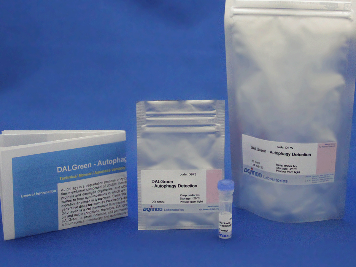
DALGreen – Autophagy Detection 细胞自噬检测试剂货号:D675
DALGreen – 细胞自噬荧光探针
DALGreen – Autophagy Detection
商品信息
储存条件:-20度保存,避光
运输条件:室温

特点:

● 操作流程简便

● 与LC3结果一致性高

● 可以动态观察细胞自噬

下载说明书
产品文献
SDS下载

选择规格:
20 nmol

操作简便

荧光/流式均可检测

自噬溶酶体检测

线粒体自噬检测(点击查看)

DALGreen &#8211; Autophagy Detection 细胞自噬检测试剂货号:D675

DALGreen &#8211; Autophagy Detection 细胞自噬检测试剂货号:D675

活动进行中
产品概述
原理
试剂概要
操作特点
实验例
常见问题Q&A
参考文献

活动进行中

订购满5000元,200元礼品等你拿

凑单关联产品TOP5

NO.1.    DAPRed – Autophagy Detection    细胞自噬检测

NO.2.    Cell Counting Kit-8     细胞增殖毒性检测   

NO.3.    Mitophagy Detection Kit    线粒体自噬检测

NO.4.    ROS Assay Kit    活性氧检测

NO.5.    Annexin V, FITC Apoptosis Detection Kit    细胞凋亡检测

产品概述

细胞自噬是细胞内损坏的蛋白质或细胞器降解和循环利用的过程。

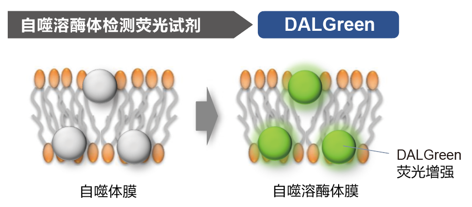
DALGreen是一种可以进入细胞膜检测细胞自噬的小分子脂溶性荧光染料,具有在酸性环境中产生荧光的特性,可以检测到自噬体与溶酶体融合形成的自噬溶酶体。

原理

DALGreen &#8211; Autophagy Detection 细胞自噬检测试剂货号:D675

DALGreen &#8211; Autophagy Detection 细胞自噬检测试剂货号:D675

与DAPGreen和DAPRed一样,在自噬体形成的时候染料掺入细胞膜。然后当自噬体与溶酶体融合产生酸性环境时,DALGreen的荧光增强。

试剂概要

DALGreen不仅可以用荧光显微镜检测,还可以使用流式细胞仪进行检测。DAPGreen可以用荧光酶标仪进行检测,

您可以根据自己的实验条件,使用不同的仪器进行检测。

DALGreen &#8211; Autophagy Detection 细胞自噬检测试剂货号:D675

操作特点

操作流程只有一步-加入试剂

无需基因转染。只需向准备好的细胞加入DALGreen染料,即可方便快捷的进行荧光检测。DALGreen &#8211; Autophagy Detection 细胞自噬检测试剂货号:D675

实验例

与LC3共染

对已经表达tagRFP-LC3的MEF细胞进行DALGreen共染实验。结果显示,DALGreen的染色部位与自噬体和自噬溶酶体的标记物LC3的位置高度一致。(比例尺:10 μm)

实验的详细情况请参考如下论文:
H. Iwashita,”Small fluorescent molecules for monitoring autophagic flux”, FEBS Letters., 2018, 592, (4), 559–567.

DALGreen &#8211; Autophagy Detection 细胞自噬检测试剂货号:D675

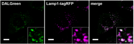
与Lamp-1共染

对已经表达Lamp1-tagRFP的MEF细胞进行DALGreen共染实验。结果显示,DALGreen的染色部位与溶酶体膜蛋白标记物Lamp 1的位置高度一致。(比例尺:10 μm)

实验的详细情况请参考如下论文:
H. Iwashita,”Small fluorescent molecules for monitoring autophagic flux”, FEBS Letters., 2018, 592, (4), 559–567.

DALGreen &#8211; Autophagy Detection 细胞自噬检测试剂货号:D675

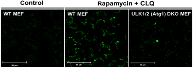
ULK1/2敲除细胞的评价


将野生型MEF细胞与已经敲除自噬体膜形成有关的ULK1/2的细胞株一起用雷帕霉素(Rapamycin)和氯喹(Chloroquine)刺激后进行对比实验。结果显示,在野生型细胞中明显观察到荧光增强,而敲除了ULK1/2的细胞中基本观察不到荧光的增强。

实验的详细情况请参考如下论文:
H. Iwashita,”Small fluorescent molecules for monitoring autophagic flux”, FEBS Letters., 2018, 592, (4), 559–567.

DALGreen &#8211; Autophagy Detection 细胞自噬检测试剂货号:D675

细胞延时成像

用DALGreen染色HeLa细胞后,按照下列条件从培养30分钟开始直至培养6小时一直对细胞染色状态进行观察。

・Nutrient Condition: 增殖型培养基
・Autophagic Condition: 不含氨基酸的培养基

<检测条件>
仪器:激光共聚焦高内涵细胞分筛系统(恒河电机株式会社: CQ1)
荧光滤光片:Ex.405 nm / Em.525/50 nm,倍率:20X

DALGreen &#8211; Autophagy Detection 细胞自噬检测试剂货号:D675

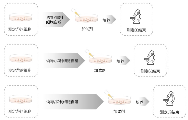
DALGreen与其它试剂的长时间染色实验的比较,可进行单一样品随时间变化的长期观察实验。
・由于DALGreen在自噬诱导/抑制剂之前加,所以可以实时观察自噬的进程。另外在不知道自噬诱导/抑制剂的作用时间情况下,与其它试剂相比,用DALGreen更加方便快捷。
每个时间组均需要单独制备样品

DALGreen &#8211; Autophagy Detection 细胞自噬检测试剂货号:D675

・MDC(Monodansylcadaverine)等其它试剂必须要在诱导后加,
需要摸索诱导/抑制剂的作用时间,因此需要准备多组实验,操作复杂,耗费更多的时间和实验材料。

与LC3的对比

DALGreen &#8211; Autophagy Detection 细胞自噬检测试剂货号:D675

HeLa细胞:①对照组,②饥饿诱导(诱导细胞自噬)组

DALGreen的荧光成像图和细胞自噬因子LC3-Ⅱ表达量结果的对比

结果
DALGreen:饥饿诱导组比Control组荧光增强;LC3-Ⅱ:饥饿诱导组比Control组LC3-Ⅱ表达量增加;
结果表明两者有良好的相关性。

自噬诱导条件
① Control:用培养基培养6小时
② 饥饿诱导:用不含氨基酸培养基培养6小时

DALGreen的显色条件
细胞:HeLa细胞
检测波长:Ex. 488 nm/Em. 500-563 nm
比例尺:20 μm

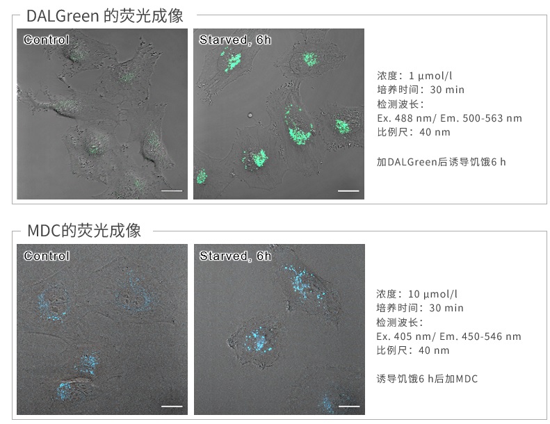
与MDC的对比

DALGreen &#8211; Autophagy Detection 细胞自噬检测试剂货号:D675

结果

DALGreen和MDC结果:饥饿诱导组均比Control组荧光增强;结果表明两者有良好的相关性。

波长

因为MDC的最大激发波长在紫外区,所以不能用488 nm激发。DALGreen可以采用488 nm激发。

操作

DALGreen:加染料⇒饥饿诱导

MDC :饥饿诱导⇒加染料

由于DALGreen是在饥饿诱导前加入,因此可以动态观察到细胞自噬的过程。

常见问题Q&A

Q1: DALGreen Working Solution的稳定性如何?
A1: 无法长期保存,需要现配现用
Q2: DMSO Stocking Solution 的稳定性如何?
A2:配制后请于-20℃保存,一个月内可保持稳定。另外建议根据用量分装保存。
Q3:如何摸索DALGreen的最佳浓度?
A3 :由于本试剂的特性,如果试剂的浓度太高或太低都会导致诱导自噬的样品组与未诱导自噬的对照组之间的差别不明显。建议参考以下信息摸索试剂的最佳浓度:

DALGreen的最佳浓度根据细胞的种类而不尽相同。

可以考虑从最低浓度(可以0.1 μmol/l作为参考)开始分别多个梯度至最高浓度(可以4 μmol/l作为参考)的步骤进行摸索。

参考例

我们公司对HeLa, HepG2, CHO细胞的最佳浓度进行了摸索。DALGreen以下列浓度进行染色,并在无氨基酸的培养基中培养以诱导自噬。下表中红色字体的浓度可明显观察到实验组与空白组的差异。

细胞种类 DALGreen浓度
HeLa 4  μmol/l 2  μmol/l 1  μmol/l 0.5  μmol/l
HepG2 4  μmol/l 2  μmol/l 1  μmol/l 0.5  μmol/l
CHO 4  μmol/l 2  μmol/l 1  μmol/l 0.5  μmol/l
Q4: 这个产品和溶酶体染色试剂有什么区别吗?
A4:溶酶体染色试剂会定位于细胞内的溶酶体里。而DALGreen是在进入自噬小体后,当自噬小体与溶酶体结合后,荧光变强。

因此当溶酶体染色试剂和DALGreen共染时,溶酶体染色试剂发出的荧光与自噬小体的部分DALGreen的荧光发生重叠。

本实验的数据,请参考下面论文的Supporting Information (Fig.S5)。

H. Iwashita, “Small fluorescent molecules for monitoring autophagic flux”, FEBS Letters., 2018, 592, (4), 559–567.

论文的原文链接在本产品的网站页面上。

网站链接(日语):

http://dominoweb.dojindo.co.jp/goodsr7.nsf/View_Display/D675?OpenDocument

Q5:推荐使用的滤光片?
A5:激发波长:350~450 nm;
发射波长:500~560 nm;
另外利用激光共聚焦显微镜的488 nm激发波长也可以检测,
请参考我们公司网站产品页面的实验例。
Q6:是否有与自噬相关的蛋白质敲除和评估的实例?
A6:与自噬小体成膜有关的ULK1和ULK2敲除的细胞与野生型细胞饥饿诱导,用DALGreen染色后评价的实例是有的。

实验结果显示,与ULK1/ULK2敲除的细胞相比,野生型细胞中的DALGreen荧光信号增大。这次实验的具体数据请参照下面这篇论文的Supporting Information (Fig. S5)

另外,自噬的标记物LC3-RFP与DALGreen的共染色结果显示,大多数的染色Puncta(斑点,位点)都高度一致。本实验的数据,请参考下面论文的Fig.1。

H. Iwashita, “Small fluorescent molecules for monitoring autophagic flux”, FEBS Letters., 2018, 592, (4), 559–567.

论文的原文链接在本产品的网站页面上。

网站链接(日语):

http://dominoweb.dojindo.co.jp/goodsr7.nsf/View_Display/D675?OpenDocument

Q7:自噬有哪些途径?DALGreen可以检测到哪些状态?
A7:众所周知,自噬根据其分子机制可以分为两种:一种是依赖于ATG5的传统自噬(LC3发生变化),另一种则是非依赖于ATG的选择性自噬(LC3形式的转化并未发生)。

DALGreen在形成自噬体膜时掺入其中,当形成自噬溶酶体时,环境变成酸性,DALGreen荧光增强。因此,DALGreen可以检测自噬溶酶体的状态。

*参考资料:发现新的自噬机制Shigeomi Shimizu

https://www.dojindo.co.jp/letterj/160/review/01.html

文献链接:http://dx.doi.org/10.14348/molcells.2018.2215

▶对于首次检测的细胞类型和实验条件,请参考FAQ[如何确定细胞自噬探针DALGreen的最佳浓度]。

Q8:在进行延时成像时有什么要注意的地方吗?
A8:为了确定最佳实验条件,请先进行预实验。

由于试剂的特性,刚刚染色后有荧光值升高的趋势,因此请参照以下步骤进行预实验和延时成像。

DALGreen &#8211; Autophagy Detection 细胞自噬检测试剂货号:D675

※条件可能因细胞种类而异

1. 预实验

– 使用对照细胞(不诱导自噬的细胞)。

– 根据说明书的步骤用Working Soluiton染色后,用培养基洗涤2次。

– 加入正常培养基后,观察荧光随时间的变化。

– 如下图所示,染色后的细胞在荧光强度阶段性降低之后,确认荧光强度变化趋于稳定的时间段(图中的T)。

※条件可能因细胞种类而异。

(参考)

HeLa细胞的话,染色后约60分钟后,荧光确定会趋于稳定(DALGreen)。

DALGreen &#8211; Autophagy Detection 细胞自噬检测试剂货号:D675

2. 延时染色成像

– 细胞用Working Solution染色后,在培养基中37℃培养。

※培养时间为预实验中摸索出的染色时间。

※染色后不要立刻进行自噬诱导。

-培养后进行自噬诱导并开始延时染色成像。

(参考)

用DALGreen对HeLa细胞进行染色,在正常的培养基中培养60分钟(预实验中摸索的时间)后,进行自噬诱导。

参考文献

No. 检测样品 检测仪器 引用(含链接)
1) 细胞
(HeLa, MEF)
荧光显微镜 H. Iwashita, H. T. Sakurai, N. Nagahora, M. Ishiyama, K. Shioji, K. Sasamoto, K. Okuma, S. Shimizu, and Y. Ueno, “Small fluorescent molecules for monitoring autophagic flux.”, FEBS Letters., 2018, 592, (4), 559–567.
2) 细胞
(HeLa)
荧光显微镜 T. Sakata, A. Saito and H. Sugimoto, “In situ measurement of autophagy under nutrient starvation based on interfacial pH sensing.”, Scientific Reports., 2018, 8, 8282.
3) 细胞
(HS-MM)
荧光显微镜 Y. Egawa, C. Saigo, Y. Kito, T. Moriki and T. Takeuchi , “Therapeutic potential of CPI-613 for targeting tumorous mitochondrial energy metabolism and inhibiting autophagy in clear cell sarcoma.”, PLoS One., 2018, 13, (6), e0198940.
4) 细胞
(HaCaT)
荧光显微镜 S. Abe, S. Hirose, M. Nishitani, I. Yoshida, M. Tsukayama, A. Tsuji and K. Yuasa , “Citrus peel polymethoxyflavones, sudachitin and nobiletin, induce distinct cellular responses in human keratinocyte HaCaT cells.”, Biosci. Biotechnol. Biochem. ., 2018, 82, (12), 1347.
5) 细胞
(KGN)
荧光显微镜 W. Yuping, M. Congshun, Z. Huihui, Z. Yuxia, C. Zhenguo and W. Liping, “Alleviation of endoplasmic reticulum stress protects against cisplatin-induced ovarian damage.”, Reprod. Biol. Endocrinol., 2018,doi: 10.1186/s12958-018-0404-4.
6) 细胞
(BmN)
荧光显微镜 S. Xue, F. Mao, D. Hu, H. Yan, J. Lei, E. Obeng, Y. Zhou, Y. Quan, and W. Yu, “Acetylation of BmAtg8 inhibits starvation-induced autophagy initiation.”, Mol. Cell Biochem., 2019,doi: 10.1007/s11010-019-03513-y.
7) 细胞
(HeLa)
荧光显微镜 F. Hongbao,Y. Shankun, C. Qixin, L. Chunyan, C. Yuqi, G. Shanshan, B. Yang, T. Zhiqi, L. Z. Amanda, T. Takanori, C.Yuncong, G. Zijian, H. Weijiang and D. Jiajie , “De Novo-Designed Near-Infrared Nanoaggregates for Super-Resolution Monitoring of Lysosomes in Cells, in Whole Organoids, and in Vivo.”, ACS Nano, 2019, 13, (12), 1446.
8) 细胞
(RT-7)
流式细胞仪 E. Sasabe, A. Tomomura, N. Kitamura and T. Yamamoto, “Metal nanoparticles-induced activation of NLRP3 inflammasome in human oral keratinocytes is a possible mechanism of oral lichenoid lesions.”, Toxicol In Vitro., 2020, 62, 104663.
9) 细胞
(骨髓细胞)
荧光显微镜 J. Xia, Y. He, B. Meng, S. Chen, J. Zhang, X. Wu, Y. Zhu, Y. Shen, X. Feng, Y. Guan, C. Kuang, J. Guo, Q. Lei, Y. Wu, G. An, G. Li, L. Qiu, F. Zhan and W. Zhou, “NEK2 induces autophagy-mediated bortezomib resistance by stabilizing Beclin-1 in multiple myeloma.”, Mol Oncol, 2020, DOI: 10.1002/1878-0261.12641.
10) 细胞
(Human L2)
荧光显微镜 Q. Xu, W. Shi, P. Lv, W. Meng, G. Mao, C. Gong, Y. Chen, Y. Wei, X. He, J. Zhao, H. Han, M. Sun and K. Xiao, “Critical role of caveolin-1 in aflatoxin B1-induced hepatotoxicity via the regulation of oxidation and autophagy.”, Cell Death Dis., 2020, 11(1), 6.
11) 细胞
(心肌细胞)
荧光显微镜 L Cui, LP Zhao, JY Ye, L Yang, Y Huang, X.P. Jiang, Q. Zhang, JZ. Jia, DX. Zhang and Y. Huang, “The Lysosomal Membrane Protein Lamp2 Alleviates Lysosomal Cell Death by Promoting Autophagic Flux in Ischemic Cardiomyocytes.”, Front Cell Dev Biol, 2020,DOI:10.3389/fcell.2020.00031.
12) 细胞
(IPEC-J2)
荧光显微镜 Y Yang, J Huang, J Li, H Yang and Y. Yin, “The Effects of Butyric Acid on the Differentiation, Proliferation, Apoptosis, and Autophagy of IPEC-J2 Cells..”, Curr. Mol. Med., 2020, 20(4), 307.
13) 细胞 (成纤维细胞、肾脏上皮细胞) 荧光显微镜 M. M. Ivanova, J. Dao, N. Kasaci, B. Adewale, J. Fikry and O. G. Alpan  , “Rapid Clathrin-Mediated Uptake of Recombinant α-Gal-A to Lysosome Activates Autophagy”, Biomolecules , 2020,  10(6), 837.
14) 细胞 (NHEKs) 荧光显微镜 S. Ikeoka and A. Kiso  , “The Involvement of Mitophagy in the Prevention of UV-B-Induced Damage in Human Epidermal Keratinocytes “, J. Soc. Cosmet. Chem. Jpn., 2020,  54(3), 252.
15) 细胞
(HeLa)
荧光显微镜(超分辨率) Q. Chen, M. Hao, L. Wang, L. Li, Y. Chen, X. Shao, Z. Tian, R. A. Pfuetzner, Q. Zhong, A. T. Brunger, J. Guan and J. Diao,”Prefused lysosomes cluster on autophagosomes regulated by VAMP8″, 2021, doi:10.1038/s41419-021-04243-0.

关联产品

DALGreen &#8211; Autophagy Detection 细胞自噬检测试剂货号:D675
线粒体自噬—Mitophagy Detection Kit
线粒体自噬检测试剂盒

DALGreen &#8211; Autophagy Detection 细胞自噬检测试剂货号:D675
DAPGreen – Autophagy Detection 细胞自噬检测试剂
DAPGreen – 细胞自噬荧光探针

DALGreen &#8211; Autophagy Detection 细胞自噬检测试剂货号:D675
DAPRed – Autophagy Detection 细胞自噬检测试剂
细胞自噬检测试剂盒

DALGreen &#8211; Autophagy Detection 细胞自噬检测试剂货号:D675
Mtphagy Dye试剂
Mtphagy Dye

DALGreen &#8211; Autophagy Detection 细胞自噬检测试剂货号:D675
LysoPrime Green – High Specificity and pH Resistance
溶酶体染色试剂Green

在线小工具

实验工具|稀释计算器|摩尔浓度计算器

CYTO-ID® 自噬检测试剂盒 CYTO-ID® Autophagy detection kit

CYTO-ID® 自噬检测试剂盒
CYTO-ID® Autophagy detection kit

  • 产品特性
  • 相关资料
  • Q&A
  • 参考文献

CYTO-ID® 自噬检测试剂盒CYTO-ID® 自噬检测试剂盒                              CYTO-ID® Autophagy detection kit

  CYTO-ID® Autophagy Detection Kit 使用新型染料检测自噬小泡和监控活细胞自噬流,选择性标记积累的自噬小泡。染料已通过优化,不染溶酶体,在自噬前体、自噬体和自噬溶酶体里呈现出明亮的荧光。该试剂盒提供了一种无需细胞转染,可以在活细胞中监控细胞自噬的快速定量方法。

CYTO-ID® 自噬检测试剂盒                              CYTO-ID® Autophagy detection kit

CYTO-ID® Autophagy Detection Kit

◆特点

● 无需转染,定量监测活细胞中的自噬情况

● 免去 LC3-GFP 转染所需的时间和精力以及转染效率的验证

● 含有特殊基团的专利染料选择性地染色自噬小泡

● 试剂盒中包含已知活性的自噬抑制剂与激活剂

● 快速量化原生异质性细胞群中的自噬

● 选择性地全方位染色,可检测和区分自噬流(autophagic flux)和自噬溶酶体的积累

● 不染溶酶体,减少其他染料的背景干扰

 便于高通量筛选自噬激活剂和抑制剂

◆原理


  该产品所用探针是阳离子两亲性示踪剂(CAT)染料,以类似诱导磷脂药物的方式迅速进入到细胞中。染料的功能基团能够选择性标记与自噬通路相关的小泡,而不会在溶酶体中聚集。


胞内物质被扩大的膜囊包裹,吞噬泡形成双层膜囊泡,成为自噬体。自噬体外膜随后与溶酶体融合,和内部物质被自噬性溶酶体降解。自噬的各种调节因子也被描绘于图中。

CYTO-ID® 自噬检测试剂盒                              CYTO-ID® Autophagy detection kit

自噬原理图

◆应用


省时省力,无需转染即可自快速全面的标记自噬小泡


为了演示 CYTO-ID® Green detection reagent 的优势,先用 RFP-LC3 表达载体转染 HeLa 细胞,10 μM 的 Tamoxifen 处理过夜,然后用 CYTO-ID® 绿色检测试剂染色。LC3 转染法需要过夜,而 CYTO-ID® 绿色检测试剂的方法在15-30分钟内标记细胞达100%。图A:绿信号显示自噬小泡的 CYTO-ID® 绿染;图B:成功转染细胞的 RFP-LC3 表达(红色);图C:自噬体的特异性标记 LC3,与 CYTO-ID® 绿色染料标记的小泡共定位的复合图像。

的发放

CYTO-ID® 自噬检测试剂盒                              CYTO-ID® Autophagy detection kit


无转染的自噬检测


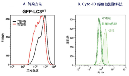
Hela细胞进行饥饿与恢复,然后使用 CYTO-ID® Green detection reagent 标记。染料可以明确检测和量化自噬及与自噬诱导相关的自噬小泡前体。图1A:稳定表达 GFP-LC3 的CHO细胞,结果显示对照组与饥饿的细胞群的相对较差的基线分离,自噬难以量化。图1B:CYTO-ID® Autophagy Detection Kit 特异性标记 LC3 蛋白依赖的自噬小泡,免去转染操作。

CYTO-ID® 自噬检测试剂盒                              CYTO-ID® Autophagy detection kit


自噬积累和自噬流的可视化


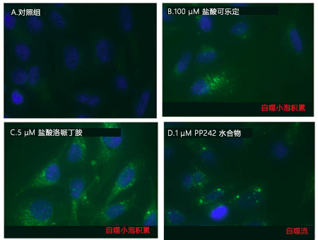
可用荧光显微镜观察CYTO-ID® 自噬绿色染料检测的自噬液泡积累和自噬流。Hela细胞用以下试剂进行模拟诱导,0.2% DMSO(A)和100 μM  Clonidine hydrochloride(B)进行诱导,5 μM Loperamide hydrochloride 和 1 μM PP242 hydrate(D)在37°C诱导12小时。处理后,细胞在37℃下用CYTO-ID® Green Detection reagent 孵育10分钟,再用分析缓冲液清洗。细胞核被 Hoechst 33342 染料复染为蓝色。

CYTO-ID® 自噬检测试剂盒                              CYTO-ID® Autophagy detection kit


避免非特异性溶酶体染色的背景干扰


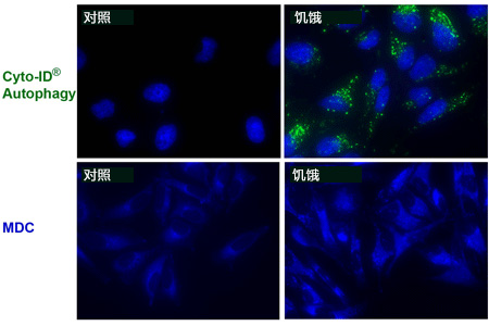
相比其他 MDC 染色溶酶体检测,CYTO-ID® 绿色染料无溶酶体染色背景,CYTO-ID® Autophagy kit 无需紫外活细胞分析,在显微镜共标记应用中,与 Hoechst 染料兼容。

CYTO-ID® 自噬检测试剂盒                              CYTO-ID® Autophagy detection kit


使用流式细胞仪通过 CYTO-ID® Autophagy Detection 

Kit 分析自噬


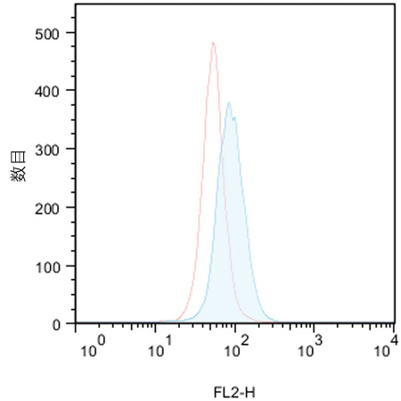
对照组(红线峰值)无诱导,10 uM Tamoxifen(ALX-550-095)处理 Jurkat 细胞(白血病T细胞)(蓝线峰值)。处理18小时后,细胞结合 CYTO-ID® 绿色检测试剂,然后不经过流式细胞仪清洗,进行分析。结果通过直方图呈现。对照组细胞被染色,但荧光亮度低。实验组中,CYTO-ID® 绿色荧光信号增加约2倍,表明 Tamoxifen 处理能引起 Jurkat 细胞中自噬的增加。


CYTO-ID® 自噬检测试剂盒                              CYTO-ID® Autophagy detection kit

CYTO-ID® 自噬检测试剂盒                              CYTO-ID® Autophagy detection kit

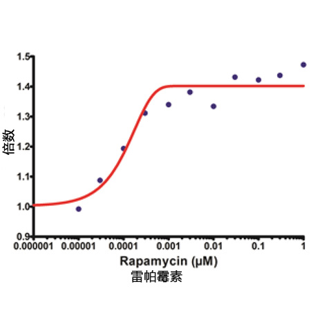
使用 mTOR 激酶抑制剂 Rapamycin 处理 HepG2 细胞孵育过夜,结果显示 CYTO-ID® 染色信号升高。

CYTO-ID® 自噬检测试剂盒                              CYTO-ID® Autophagy detection kit

CYTO-ID® 绿色染料大多与 RFP-LC3蛋白共定位。0.1 μM Rapamycin(典型自噬诱导剂)处理过夜,转染 Hela 细胞表达RFP-LC3。

A: CYTO-ID® Green 染色; B: RFP-LC3; C: 合成图像。

CYTO-ID® 自噬检测试剂盒 2.0

◆相关产品

产品编号

产品名称

规格

备注

应用

ENZ-51002-25

GFP-Certified®Apoptosis/Necrosis detection kit

细胞凋亡/坏死检测试剂盒

25 assays

多重检测,区分正常、

早期凋亡、晚期凋亡和坏死细胞,与GFP和其他绿色荧光探针兼容。

FC,荧光显微镜,荧光检测

ENZ-51002-100

100 assays

ENZ-51021-K200

Nuclear-ID®Green

hromatin condensation detection kit

细胞核绿色染色体皱缩检测试剂盒


1 Kit

高渗透性的绿色荧光染色增强了细胞凋亡诱导染色质固缩。


FC,荧光显微镜,荧光检测

ENZ-52406

NUCLEAR-ID®Red DNA stain

DNA染色试剂盒(红色荧光)

200 µL

细胞可渗透的DNA染色应用广泛。

≥93%(HPLC),FC,荧光检测

ENZ-CHM103-0200

Nuclear-ID® Blue

DNA stain   (GFP-Certified®)

Nuclear ID® 蓝色DNA染色(GFP细胞系)

200 µL

细胞可渗透的DNA染色应用广泛。

≥93%(HPLC),FC,荧光检测

ENZ-51015-KP002

Lyso-ID® Red

cytotoxicity kit   (GFP-Certified®)

溶酶体细胞毒理检测试剂盒(红色荧光)(绿色细胞系)

1 Kit

快速,定量和HTS-兼容的检测活细胞毒性试剂。

荧光显微镜,荧光检测

ENZ-51035-0025

PROTEOSTAT®Aggresome   etection kit

蛋白内稳态聚集体 检测 试剂盒

25 tests

Robust、定量的聚集小体用于神经退行性疾病,肝病和毒理学研究

FC,荧光显微镜,荧光检测

ENZ-51035-K100

100 tests


处理

目的

效果

μM

诱导时间 (hrs)

细胞系

饥饿

抑制哺乳动物Rapamycin (mTOR)

激活自噬

N/A

1~4

HeLa, HepG2, Jurkat

Rapamycin

抑制哺乳动物Rapamycin (mTOR)

激活自噬

0.2

6~18

HeLa, Jurkat

PP242

mTOR ATP-竞争性抑制剂

激活自噬

1

18

HeLa

Lithium

抑制IMPase 和降低inositol 和IP3水平; mTOR依赖性

激活自噬

10,000

18

HeLa, Jurkat

Trehalose

未知, mTOR依赖性

激活自噬

50,000

6

HeLa, Jurkat

Bafilomycin A1

抑制 Vacuolar-ATPase

抑制自噬

6~9*10-3

18

HeLa, Jurkat

Chloroquine

碱化 Lysosomal pH

抑制自噬

10~50

18

HeLa, Jurkat

Tamoxifen

增加细胞内 ceramide的水平和消除PI3K的抑制效果

激活自噬

4~10

6~18

HeLa, HepG2, Jurkat

Verapamil

钙离子通道阻滞剂;降低胞浆内Ca2+水平; mTOR依赖性

激活自噬

40~100

18

HeLa, Jurkat

HydroxyChloroquine

Alkalinizes Lysosomal pH

抑制自噬 

10

18

HeLa, Jurkat

Loperamide

Ca2+ 通道阻滞剂;降低胞浆内 Ca2+浓度; mTOR依赖性

激活自噬

5

18

HeLa

Clonidine

咪唑啉-1受体激动剂; 降低 cAMP 水平; mTOR依赖性

激活自噬

100

18

HeLa

MG-132

选择性蛋白酶抑制剂

激活自噬

2~5

18

HeLa, Jurkat

Norclomipramine

碱化Lysosomal pH

抑制自噬

5-20

18

HeLa

Epoxomicin

选择性蛋白酶抑制剂

诱导聚集体

0.5

18

HeLa

Velcade®

选择性蛋白酶抑制剂

诱导聚集体

0.5

18

HeLa

Amyloid beta peptide 1-42

引起氧化应激

诱导聚集体

25

18

SK-N-SH

Cyto-ID®检测试剂盒 Q&A


Q:Cyto-ID®自噬检测试剂与活细胞孵育的最长时间是多少(几分钟/几小时/几天)?
A:我们建议 37℃ 孵育不超过 30 分钟。很多实验表明,孵育 1 小时不会伤害细胞。孵育超过 1 小时,一些细胞看上去状态不好。


Q:是否可用荧光显微镜观察 3D 培养细胞的自噬?
A:我们没有用该染料染过 3D 细胞,但是一小撮细胞是可以染色的。活体组织的话,由于染料的渗透性不好,不能进入样本的内部,所以效果不

A:想。染白细胞是可以。


Q:用双重染料染在 37℃ 孵育 30 分钟后,细胞状态不好开始皱缩。有些细胞也从板上脱离,阳性对照和 DMSO 对照没有观察到特异性染色。
A:对于比较敏感的细胞,优化流程如下:
A:1. 使用 PBS/5% FBS 或者完全培养基作为 assay buffer,最好不含酚红。
A:2. 如果细胞状态仍然不好,建议染色时间缩短至 15~20 分钟。
A:3. 减少染料的浓度至 1000× 稀释。需要显微镜观察时延长荧光照射的时间。
A:4. 可能光褪色会导致信号模糊。建议使用封片剂防止光褪色。


Q:用流式检测的话,样品在用 Cyto-ID®自噬检测试剂孵育后多久可用流式观察?操作手册是说 30 分钟。是否可以在检测之前短暂的将样品放

A:在冰上?
A:可以将样品放在冰上延长孵育的时间。一般的经验法则,尽可能缩短活细胞与染料的结合。缺氧,低温或者其他应激状态也可以诱导自噬。


Q:GFP-CertifiedTM 细胞凋亡/坏死检测试剂盒(ENZ-51002),Cyto-ID® 自噬检测试剂盒(ENZ-51031)是否可以同时染色,检测细胞凋

A:亡、坏死和自噬?
A:GFP-CertifiedTM 细胞凋亡/坏死检测试剂盒(ENZ-51002)和 Cyto-ID® 检测试剂盒(ENZ-51031)不能配合使用,因为细胞凋亡和自噬检

A:测的染料都发绿色荧光。这两个试剂盒可以平行使用。比如,用十字孢碱(Staurosporine)处理细胞,一部分样品用 GFP-CertifiedTM 细胞

A:凋亡/坏死检测试剂盒检测,一部分样品用 Cyto-ID® 检测试剂盒检测。


Q:Cyto-ID®自噬检测试剂盒(ENZ-51031)的 Cyto-ID®自噬检测试剂能否与 ProteoStat®蛋白聚集检测(ENZ-51023)的 ProteoStat®

A:检测试剂联合使用?
A:Cyto-ID®绿色自噬检测试剂与 ProteoStat®检测试剂发的荧光颜色不同,他们可用于同一实验,客户需要优化实验流程。

Q:Cyto-ID®自噬检测试剂的原理是什么?
A:染料是申请专利的。该染料是阳离子两性示踪剂(CAT)染料,跟很多阳离子化合物一样可以快速进入细胞。染料穿过细胞膜双分子层后被动

A:扩散,不需要蛋白结合或者转运子活性。可滴定部分的筛选使的染料不会再溶酶体聚集,能够标记自噬通道相关的液泡。另外,染料的发射光

A:密度增强后可以标记自噬液泡相关片层膜结构。我们可以提供染料染自噬液泡的相关数据。诱导自噬溶酶体,比如用氯喹或者baflimyocin 处

A:理后,会导致荧光变亮(250~300%)。雷帕霉素诱导自噬后能够用3-MA 抑制。在EBSS 中氨基酸缺乏会导致1 小时后在自噬液泡中染料的

A:积累,这种积累是可逆的(补充营养后即可恢复)。我们有一系列用于研究自噬的化合物(Screen-Well®Autophagy library)


Q:与单丹磺酰尸胺(MDC)相比,Cyto-ID®自噬检测试剂有哪些优势?
A:MDC 需要 365 nm UV 荧光照明,与通常流式配置的 488 nm 激发源不兼容。Cyto-ID®自噬染料是 488 nm 激发波长,绿色的发射荧光,可

A:以高亮自噬通道不同的液泡成分。需要注意的是,与 lysomotrophic 染料不同,MDC、LysoTrackerTMRed、吖啶橙主要是染溶酶体的,

A:Cyto-ID®自噬染料只能染一点点溶酶体,主要是染自噬溶酶体和早期自噬部分的选择标记物。


Q:与 GFP-LC3 转染细胞相比,Cyto-ID®自噬检测试剂盒(ENZ-51031)的优势是什么?
A:Cyto-ID®检测试剂盒(ENZ-51031)的优势在于不用转染细胞。转染很短暂,不同实验需要再购买相关试剂。由于细胞过表达,会导致

A:GFP-LC3 聚集,结果呈假阳性。转染效率低于100%,有些细胞不会表达必要的 GFP 组件。该试剂盒检测的自噬,细胞群的一致性会更好。

A:不需要每个细胞系都进行转染。也可以标记原代细胞。原代细胞不容易进行转染。


Q:有哪些细胞系或者原代细胞可用 Cyto-ID®自噬检测试剂?
A:用该试剂盒见检测过的代表性的细胞和细胞系包括肝细胞、SK-N-SH 神经瘤细胞、CHO 细胞、骨肉瘤来源细胞、黑素留细胞、乳腺癌细胞、

A:宫颈癌细胞、卵巢癌细胞、B 淋巴瘤细胞、结肠癌细胞、HepG 细胞、Jurkat T细胞、牛大动脉内皮细胞(BAEC)。


Q:Cyto-ID®自噬检测试剂能否与其他染料配合使用,区分细胞核内体和自噬内涵体?
A:Cyto-ID®红色长期示踪剂染料(ENZ-51037)能够染细胞质膜,通过细胞内吞存在于细胞中。Cyto-ID®红色染料与 Cyto-ID®自噬检测试剂

A:能够高亮异体吞噬 vs 自噬。也可以 Cyto-ID®自噬检测试剂与Lyso-ID®红色染料或者LysoTrackerTM 红色染料进行活细胞染色,荧光显微镜

A:观察。


Q:Cyto-ID®自噬检测试剂的稳定性如何?
A:荧光染料会发生光褪色,但是这种染料比荧光素更稳定。按照操作手册出来荧光染料。

Q:Cyto-ID®自噬检测试剂盒染色后是否可固定细胞?
A:在操作手册中提供了固定细胞的流程。不推荐用表面活性剂给细胞打孔,定位的荧光染料丢失。


Q:活细胞检测时,荧光信号强度可维持多长时间?
A:细胞诱导后发生自噬,在分析前染色。诱导时间少于 1 小时或者诱导几天,诱导时间取决于自噬诱导剂。我们没有在活细胞上进行长时间的染

A:色,有客户发现可以至少染色 24 小时。


参考文献


1.

A comparison of strategies for immortalizing mouse embryonic fibroblasts: M.M. St. Amad, et al.; J. Biol. Methods 3, e41 (2016), Application(s): Detected autophagy induction in SV40 transformed versus serially passed MEFs, 全文

2.

Astemizole-Histamine induces Beclin-1-independent autophagy by targeting p53-dependent crosstalk between autophagy and apoptosis: R. Jakhar, et al.; Cancer Lett. 372, 89 (2016), Application(s): Flow cytometry analysis, 摘要;

3.

Atg5 Is Essential for the Development and Survival of Innate Lymphocytes: T.E. O'Sullivan, et al.; Cell Rep. 15, 1910 (2016), Application(s): Liver lymphocytes were harvested and stained with cell surface antibodies and then incubated with 1:400 Cyto-ID Autophagy Detection Reagent, 摘要全文

4.

Autophagy-related gene 5 and Wnt5 signaling pathway requires differentiation of embryonic stem cells into odontoblast-like cells: N. Ozeki, et al.; Exp. Cell Res. 341, 92 (2016), Application(s): Autophagy flux, 摘要;

5.

DHA-induced stress response in human colon cancer cells-focus on oxidative stress and autophagy: K. Pettersen, et al.; Free Radic. Biol. Med. 90, 158 (2016), Application(s):Autophagy determined by flow cytometry, 摘要;

6.

Diosgenin induces ROS-dependent autophagy and cytotoxicity via mTOR signaling pathway in chronic myeloid leukemia cells: S. Jiang, et al.; Phytomedicine 23, 243 (2016),Application(s): Confocal immunofluorescence, 摘要;

7.

Efavirenz causes oxidative stress, endoplasmic reticulum stress, and autophagy in endothelial cells: M. Weiss, et al.; Cardiovasc. Toxicol. 16, 90 (2016), 摘要;

8.

MiR-193b promotes autophagy and non-apoptotic cell death in oesophageal cancer cells: M.J. Nyhan, et al.; BMC Cancer 16, 101 (2016), Application(s): Assay used to stain live cells, 摘要全文

9.

(Pro)renin receptor regulates autophagy and apoptosis in podocytes exposed to high glucose: C. Li, et al.; Am. J. Physiol. Endocrinol. Metab. 309, E302 (2015), Application(s):Confocal microscopy using mouse podocytes, 摘要;

10.

A rapid and high content assay that measures cyto-ID-stained autophagic compartments and estimates autophagy flux with potential clinical applications: S. Guo, et al.; Autophagy11, 560 (2015), Application(s): Fluorescent detection, Microplate Reader , 摘要全文

11.

A Systems Approach Identifies Essential FOXO3 Functions at Key Steps of Terminal Erythropoiesis: R. Liang, et al.; PLoS Genet. 11, e1005526 (2015), Application(s):Autophagy flux measured by flow cytometry , 摘要全文

12.

ABT-888 enhances cytotoxic effects of temozolomide independent of MGMT status in serum free cultured glioma cells: R.K. Balvers, et al.; J. Transl. Med. 13, 74 (2015),Application(s): Assay, 摘要全文

13.

Activation of autophagy in response to nanosecond pulsed electric field exposure: J.C. Ullery, et al.; Biochem. Biophys. Res. Commun. 458, 411 (2015), Application(s):Fluorescence microscopy using U937 monocyte and CHO-K1 cell lines, 摘要;

14.

Aflatoxin biosynthesis is a novel source of reactive oxygen species-a potential redox signal to initiate resistance to oxidative stress?: L.V. Roze, et al.; Toxins (Basel). 7, 1411 (2015),Application(s): Assay, 摘要全文

15.

Alisertib induces cell cycle arrest and autophagy and suppresses epithelial-to-mesenchymal transition involving PI3K/Akt/mTOR and sirtuin 1-mediated signaling pathways in human pancreatic cancer cells: F. Wang, et al.; Drug Des. Devel. Ther. 9, 575 (2015), Application(s): Flow cytometry using PANC-1 and BxPC-3 pancreatic cancer cell lines, 摘要全文

16.

Alisertib, an Aurora kinase A inhibitor, induces apoptosis and autophagy but inhibits epithelial to mesenchymal transition in human epithelial ovarian cancer: Y.H. Ding, et al.; Drug Des. Devel. Ther. 9, 425 (2015), Application(s): Flow cytometry using SKOV3 and OVCAR-4 ovarian cancer cell lines, 摘要全文

17.

Andrographolide Analogue Induces Apoptosis and Autophagy Mediated Cell Death in U937 Cells by Inhibition of PI3K/Akt/mTOR Pathway: D. Kumar, et al.; PLoS One 10, e0139657 (2015), Application(s): Flow cytometric analysis of Cyto-ID Green Detection Reagent , 摘要全文

18.

Apoptotic Cell Death Induced by Resveratrol Is Partially Mediated by the Autophagy Pathway in Human Ovarian Cancer Cells: F. Lang, et al.; PLoS One 10, e0129196 (2015),Application(s): Live Cell Imaging, 摘要全文

19.

Araguspongine C induces autophagic death in breast cancer cells through suppression of c-Met and HER2 receptor tyrosine kinase signaling: M.R. Akl, et al.; Mar. Drugs 13, 288 (2015), Application(s): Flow cytometry using BT-474 breast cancer cell line, 摘要全文

20.

Autocrine VEGF maintains endothelial survival through regulation of metabolism and autophagy: C.K. Domigan, et al.; J. Cell. Sci. 128, 2236 (2015), 摘要;

21.

Autophagy is activated in systemic lupus erythematosus and required for plasmablast development: A.J. Clarke, et al.; Ann. Rheum. Dis. 74, 912 (2015), 摘要全文

22.

Autophagy limits proliferation and glycolytic metabolism in acute myeloid leukemia: A.S. Watson, et al.; Cell Death Discov. 1, 15008 (2015), Application(s): CytoID assay in human and mouse HSCs, 摘要全文

23.

Baicalin inhibits autophagy induced by influenza A virus H3N2: H.Y. Zhu, et al.; Antiviral Res. 113, 62 (2015), Application(s): Fluorescence microscopy using A549 human lung cancer cell line, 摘要;


24.

Bardoxolone methyl induces apoptosis and autophagy and inhibits epithelial-to-mesenchymal transition and stemness in esophageal squamous cancer cells: Y.Y. Wang, et al.; Drug Des. Devel. Ther. 9, 993 (2015), Application(s): Flow Cytometry, 摘要全文

25.

Cell-penetrating peptide derived from human eosinophil cationic protein inhibits mite allergen Der p 2 induced inflammasome activation: S.J. Yu, et al.; PLoS One 10, e0121393 (2015), Application(s): Flow cytometry of THP-1 leukemia cell line, 摘要全文

26.

Chemoproteomics Reveals Novel Protein and Lipid Kinase Targets of Clinical CDK4/6 Inhibitors in Lung Cancer: N.J. Sumi, et al.; ACS Chem. Biol. 10, 2680 (2015),Application(s): Quantification of autophagosomes, 摘要;

27.

Circulating hemocytes from larvae of the Japanese rhinoceros beetle Allomyrina dichotoma (Linnaeus) (Coleoptera: Scarabaeidae) and the cellular immune response to microorganisms: S. Hwang, et al.; PLoS One 10, e0128519 (2015), Application(s):Fluorescence microscopy using hemocytes from Japanese rhinoceros beetle Allomyrina dichotoma larvae, 摘要全文

28.

Citreoviridin induces ROS-dependent autophagic cell death in human liver HepG2 cells: Y.N. Liu, et al.; Toxicon. 95, 30 (2015), Application(s): Fluorescence microscopy using HepG2 cell line, 摘要;

29.

Clozapine induces autophagic cell death in non-small cell lung cancer cells: Y.C. Yin, et al.; Cell. Physiol. Biochem. 35, 945 (2015), 摘要;

30.

Coffee and caffeine potentiate the antiamyloidogenic activity of melatonin via inhibition of Aβ oligomerization and modulation of the Tau-mediated pathway in N2a/APP cells: L.F. Zhang, et al.; Drug Des. Devel. Ther. 9, 241 (2015), Application(s): Flow Cytometry,摘要全文

31.

Combination of the mTOR inhibitor RAD001 with temozolomide and radiation effectively inhibits the growth of glioblastoma cells in culture: H. Burckel, et al.; Oncol. Rep. 33, 471 (2015), 摘要;

32.

Danusertib Induces Apoptosis, Cell Cycle Arrest, and Autophagy but Inhibits Epithelial to Mesenchymal Transition Involving PI3K/Akt/mTOR Signaling Pathway in Human Ovarian Cancer Cells: D. Zi, et al.; Int. J. Mol. Sci. 16, 27228 (2015), Application(s): Confocal fluorescence microscopy, 摘要全文

33.

Danusertib, a potent pan-Aurora kinase and ABL kinase inhibitor, induces cell cycle arrest and programmed cell death and inhibits epithelial to mesenchymal transition involving the PI3K/Akt/mTOR-mediated signaling pathway in human gastric cancer AGS and NCI-N78 cells: C.X. Yuan, et al.; Drug Des. Devel. Ther. 9, 1293 (2015), Application(s): Flow cytometry using AGS and NCI-N78 gastric cancer cell lines, 摘要全文

34.

Defective autophagy in vascular smooth muscle cells alters contractility and Ca²⁺ homeostasis in mice: C.F. Michiels, et al.; Am. J. Physiol. Heart Circ. Physiol. 308, H557 (2015), 摘要;

35.

Effects of cyclodextrins on GM1‐gangliosides in fibroblasts from GM1‐gangliosidosis patients: Y. Maeda, et al.; J. Pharm. Pharmacol. 67, 1133 (2015), 摘要;

36.

Endurance exercise training induces fat depot-specific differences in basal autophagic activity: G. Tanaka, et al.; Biochem. Biophys. Res. Commun. 466, 512 (2015),Application(s): Detect the formation of autophagosomes, 摘要;

37.

Erbin is a novel substrate of the Sag-βTrCP E3 ligase that regulates KrasG12D-induced skin tumorigenesis: C.M. Xie, et al.; J. Cell. Biol. 209, 721 (2015), 摘要;

38.

Evaluation of Antitumor Effects of Folate-Conjugated Methyl-β-cyclodextrin in Melanoma: K. Motoyama, et al.; Biol. Pharm. Bull. 38, 374 (2015), Application(s): Fluorescence Microscopy, 摘要全文

39.

Exchange protein directly activated by cAMP 1 promotes autophagy during cardiomyocyte hypertrophy: A.C. Laurent, et al.; Cardiovasc. Res. 105, 55 (2015), Application(s):Fluorescence microscopy using rat neonatal ventricular myocytes, 摘要;

40.

Glutathione-S-transferase omega 1 (GSTO1-1) acts as mediator of signaling pathways involved in aflatoxin B1-induced apoptosis-autophagy crosstalk in macrophages: S. Paul, et al.; Free Radic. Biol. Med. 89, 1218 (2015), Application(s): Determination of autophagy with immunocytochemistry , 摘要;

41.

GMI, an immunomodulatory protein from Ganoderma microsporum, potentiates cisplatin-induced apoptosis via autophagy in lung cancer cells: I.L. Hsin, et al.; Mol. Pharm. 12, 1534 (2015), 摘要;

42.

Induction of apoptosis and autophagy via sirtuin1- and PI3K/Akt/mTOR-mediated pathways by plumbagin in human prostate cancer cells: Z.W. Zhou, et al.; Drug Des. Devel. Ther. 9, 1511 (2015), Application(s): Assay, 摘要全文

43.

Induction of autophagy is a key component of all-trans-retinoic acid-induced differentiation in leukemia cells and a potential target for pharmacologic modulation: N. Orfali, et al.; Exp. Hematol. 43, 781 (2015), Application(s): Flow cytometry analysis of NB4 and HL60 promyelocytic leukemia cell lines, 摘要;

44.

Inhibition of Autophagy Potentiated the Antitumor Effect of Nedaplatin in Cisplatin-Resistant Nasopharyngeal Carcinoma Cells: Z. Liu, et al. ; PLoS One 10, e0135236 (2015),Application(s): Cell culture, 摘要全文

45.

Inhibition of mitotic Aurora kinase A by alisertib induces apoptosis and autophagy of human gastric cancer AGS and NCI-N78 cells: C.X. Yuan, et al.; Drug Des. Devel. Ther. 9, 487 (2015), Application(s): Flow cytometry using AGS and NCI-N78 gastric cancer cell lines, 摘要全文

46.

Interferon Regulatory Factor-1 signaling regulates the switch between autophagy and apoptosis to determine breast cancer cell fate: J.L. Schwartz-Roberts, et al.; Cancer Res.75, 1046 (2015), 摘要;

47.

Interplay of Oxidative Stress and Autophagy in PAMAM Dendrimers-Induced Neuronal Cell Death : Y. Li, et al.; Theranostics 5, 1363 (2015), Application(s): Confocal fluorescence assay, 摘要全文

48.

Invariant NKT cells require autophagy to coordinate proliferation and survival signals during differentiation: B. Pei, et al.; J. Immunol. 194, 5872 (2015), 摘要;

49.

Involvement of fish signal transducer and activator of transcription 3 (STAT3) in nodavirus infection induced cell death: Y. Huang, et al.; Fish Shellfish Immunol. 43, 241 (2015),Application(s): Fluorescence microscopy of Grouper (fish) brain cells, 摘要;

50.

Is the autophagy a friend or foe in the silver nanoparticles associated radiotherapy for glioma?: H. Wu, et al.; Biomaterials 62, 47 (2015), Application(s): Fluorescence microscopy using U251 human glioma cell line, 摘要;

51.

Kaposi's sarcoma-associated herpesvirus induces Nrf2 activation in latently infected endothelial cells through SQSTM1 phosphorylation and interaction with polyubiquitinated Keap1: O. Gjyshi, et al.; J. Virol. 89, 2268 (2015), 摘要;

52.

KLF4-SQSTM1/p62-associated prosurvival autophagy contributes to carfilzomib resistance in multiple myeloma models: I. Riz, et al.; Oncotarget 6, 17814 (2015), Application(s):FACS, 摘要全文

53.

Lithium modulates autophagy in esophageal and colorectal cancer cells and enhances the efficacy of therapeutic agents in vitro and in vivo: T.R. O'Donovan, et al.; PLoS One 10, e0134676 (2015), Application(s): Flow cytometry analysis using human esophageal and murine colon cancer cell lines, 摘要全文

54.

Methicillin-Resistant Staphylococcus aureus Adaptation to Human Keratinocytes: G. Soong, et al.; MBio. 6, e00289-15 (2015), Application(s): Assay, 摘要全文

55.

Mevalonate pathway regulates cell size homeostasis and proteostasis through autophagy: T.P. Miettinen, et al.; Cell Rep. 13, 2610 (2015), Application(s): Flow cytometry analysis of autophagy using Jurkat, U2OS, Kc167 and HUVEC cells, 摘要;

56.

MiR-29b replacement inhibits proteasomes and disrupts aggresome+autophagosome formation to enhance the antimyeloma benefit of bortezomib: S. Jagannathan, et al.; Leukemia 29, 727 (2015), Application(s): Detection of autophagy by fluorescence microscopy in multiple myeloma cell lines, 摘要全文

57.

Molecular chaperone GRP78 enhances aggresome delivery to autophagosomes to promote drug resistance in multiple myeloma: M.A. Abdel Malek, et al.; Oncotarget 6, 3098 (2015), Application(s): Confocal Microscopy, 摘要全文

58.

Molecular cloning and characterization of autophagy-related gene TmATG8 in Listeria-invaded hemocytes of Tenebrio molitor: H. Tindwa, et al.; Dev. Comp. Immunol. 51, 88 (2015), Application(s): Fluorescence microscopy using hemocytes from Tenebrio molitor larvae, 摘要;

59.

Molecular pathway of near-infrared laser phototoxicity involves ATF-4 orchestrated ER stress: I. Khan, et al.; Sci. Rep. 5, 10581 (2015), Application(s): Fluorescence microscopy autophagy assay, 摘要全文

60.

N-Myc and STAT Interactor regulates autophagy and chemosensitivity in breast cancer cells: B.J. Metge, et al.; Sci. Rep. 5, 11995 (2015), Application(s): Fluorescent detection,摘要全文

61.

Novel autophagy inducers lentztrehaloses A, B and C: S.I. Wada, et al.; J. Antibiot. (Tokyo)68, 521 (2015), Application(s): Fluorescence microscopy using Mewo melanoma and OVK18 ovarian cancer cell lines, 摘要;

62.

Novel small-molecule SIRT1 inhibitors induce cell death in adult T-cell leukaemia cells: T. Kozako, et al.; Sci. Rep. 5, 11345 (2015), Application(s): Flow cytometry using a variety of cancer cell lines, 摘要全文

63.

Novel targeting of PEGylated liposomes for codelivery of TGF-β1 siRNA and four antitubercular drugs to human macrophages for the treatment of mycobacterial infection: a quantitative proteomic study: N. Niu, et al. ; Drug Des. Devel. Ther. 9, 4441 (2015),Application(s): Autophagy of human macrophages by flow cytometry, 摘要;

64.

Paraptosis cell death induction by the thiamine analog benfotiamine in leukemia cells: N. Sugimori, et al.; PLoS One 10, e0120709 (2015), Application(s): Flow cytometry using HL60 leukemia cell line, 摘要全文

65.

Plumbagin induces G2/M arrest, apoptosis, and autophagy via p38 MAPK- and PI3K/Akt/mTOR-mediated pathways in human tongue squamous cell carcinoma cells: S.T. Pan, et al.; Drug Des. Devel. Ther. 9, 1601 (2015), Application(s): Assay, 摘要全文

66.

Pro-apoptotic and pro-autophagic effects of the Aurora kinase A inhibitor alisertib (MLN8237) on human osteosarcoma U-2 OS and MG-63 cells through the activation of mitochondria-mediated pathway and inhibition of p38 MAPK/PI3K/Akt/mTOR signaling pathway: N.K. Niu, et al.; Drug Des. Devel. Ther. 9, 1555 (2015), Application(s): Assay,摘要全文

67.

Reduced FoxO3a expression causes low autophagy in idiopathic pulmonary fibrosis fibroblasts on collagen matrix: J. Im, et al.; Am. J. Physiol. Lung Cell. Mol. Physiol. 309, L552 (2015), 摘要;

68.

S-Adenosyl-L-methionine-competitive inhibitors of the histone methyltransferase EZH2 induce autophagy and enhance drug sensitivity in cancer cells: T.P. Liu, et al.; Anticancer Drugs 26, 139 (2015), Application(s): Fluorescence microscopy using MDA-MB-231 breast cancer cell line, 摘要全文

69.

Schisandrin B inhibits cell growth and induces cellular apoptosis and autophagy in mouse hepatocytes and macrophages: implications for its hepatotoxicity: Y. Zhang, et al.; Drug Des. Devel. Ther. 9, 2001 (2015), Application(s): Flow cytometry using AML-12 hepatocyte and RAW 264.7 leukemia cell lines, 摘要全文

70.

Src/STAT3-dependent heme oxygenase-1 induction mediates chemoresistance of breast cancer cells to doxorubicin by promoting autophagy: Q. Tan, et al.; Cancer Sci. 106, 1023 (2015), 摘要;

71.

The CCL2 chemokine is a negative regulator of autophagy and necrosis in luminal B breast cancer cells: W.B. Fang, et al.; Breast Cancer Res. Treat. 150, 309 (2015), 摘要;

72.

The investigational Aurora kinase A inhibitor alisertib (MLN8237) induces cell cycle G2/M arrest, apoptosis, and autophagy via p38 MAPK and Akt/mTOR signaling pathways in human breast cancer cells: J.P. Li, et al.; Drug Des. Devel. Ther. 9, 1627 (2015),Application(s): Assay, 摘要全文

73.

The pan-inhibitor of Aurora kinases danusertib induces apoptosis and autophagy and suppresses epithelial-to-mesenchymal transition in human breast cancer cells: J.P. Li, et al.; Drug Des. Devel. Ther. 9, 1027 (2015), Application(s): Assay, 摘要全文

74.

The role of autophagy in the cytotoxicity induced by recombinant human arginase in laryngeal squamous cell carcinoma: C. Lin, et al.; Appl. Microbiol. Biotechnol. 99, 8487 (2015), 摘要;

75.

A Novel CXCR3-B Chemokine Receptor-induced Growth-inhibitory Signal in Cancer Cells Is Mediated through the Regulation of Bach-1 Protein and Nrf2 Protein Nuclear Translocation : M. Balan & S. Pal; J. Biol. Chem. 289, 3126 (2014), Application(s): Monitor autophagy in MCF-7 and T47D breast cancer cells by flow cytometry and fluorescence microscopy, 摘要;

76.

Adaptive responses to glucose restriction enhance cell survival, antioxidant capability, and autophagy of the protozoan parasite Trichomonas vaginalis: K.Y. Huang, et al.; Biochim. Biophys. Acta. 1840, 53 (2014), 摘要;

77.

Autophagy in the brain of neonates following hypoxia-ischemia shows sex-and region-specific effects: S.N. Weis, et al.; Neuroscience 256, 201 (2014), 摘要;

78.

Cannabinoid-induced autophagy regulates suppressor of cytokine signaling-3 in intestinal epithelium: L.C. Koay, et al.; Am. J. Physiol. Gastrointest. Liver Physiol. 307, G140 (2014),Application(s): Detection of autophagy in human colonic epithelial cell line Caco-2 by Confocal imaging, 摘要全文

79.

Caveolin-1 Is a Critical Determinant of Autophagy, Metabolic Switching, and Oxidative Stress in Vascular Endothelium: T. Shiroto, et al.; PLoS One 9, e87871 (2014), 摘要;全文

80.

Connective tissue diseases: How do autoreactive B cells survive in SLE-autophagy?: N.J. Bernard; Nat. Rev. Rheumatol. 10, 128 (2014), (Review), 摘要;

81.

Defective Autophagosome Trafficking Contributes to Impaired Autophagic Flux in Coronary Arterial Myocytes Lacking CD38 Gene: Y. Zhang, et al.; Cardiovasc. Res. 102, 68 (2014),摘要;

82.

Defects in mitochondrial clearance predispose human monocytes to interleukin-1β hyper-secretion: R. van der Burgh, et al.; J. Biol. Chem. 289, 5000 (2014), 摘要全文

83.

Early biomarkers of response to carfilzomib in multiple myeloma (MM): Modulation of CXCR4 and induction of autophagy: M. Bhutani, et al.; J. Clin. Oncol. 32, e19572 (2014),Application(s): Quantification of autophagy in malignant plasma cells from bone marrow aspirates by flow cytometry with the Cyto-ID autophagy detection kit,

84.

Enhancement of dynein-mediated autophagosome trafficking and autophagy maturation by ROS in mouse coronary arterial myocytes: M. Xu, et al.; J. Cell. Mol. Med. 18, 2165 (2014), 摘要全文

85.

Flow Cytometric Analysis of Autophagic Activity with Cyto-ID Staining in Primary Cells: M. Stankov, et al.; Bio-Protocol (2014), Application(s): FC in primary BMDCs, 全文

86.

High-Content Assays for Hepatotoxicity Using Induced Pluripotent Stem Cell-Derived Cells: O. Sirenko, et al.; Assay Drug Dev. Technol. 12, 43 (2014), 摘要全文

87.

Histone deacetylase inhibitors induce apoptosis in myeloid leukemia by suppressing autophagy: M.V. Stankov, et al.; Leukemia 28, 577 (2014), 摘要;

88.

Histone deacetylase inhibitors potentiate VSV oncolysis in prostate cancer cells by modulating NF-κB dependent autophagy: L. Shulak, et al.; J. Virol. 88, 2927 (2014),摘要;

89.

In vitro and in vivo characterization of porcine acellular dermal matrix for gingival augmentation procedures: A.M. Pabst, et al.; J. Periodontal. Res. 49, 371 (2014), 摘要;

90.

Inhibition of Autophagic Flux by Salinomycin Results in Anti-Cancer Effect in Hepatocellular Carcinoma Cells: J. Klose, et al.; PLoS One 9, e95970 (2014),Application(s): Autophagy detection in human hepatocellular carcinoma , 摘要全文

91.

Inhibition of stress induced premature senescence in presenilin-1 mutated cells with water soluble Coenzyme Q10: D. Ma, et al.; Mitochondrion 17C, 106 (2014), Application(s):Autophagic vacuoles in Alzheimer's Disease fibroblasts detected with CytoID® Green Autophagy Detection kit, 摘要;

92.

Involvement of autophagy in recombinant human arginase-induced cell apoptosis and growth inhibition of malignant melanoma cells: Z. Wang, et al.; Appl. Microbiol. Biotechnol.98, 2485 (2014), 摘要;

93.

MiR-216a: a link between endothelial dysfunction and autophagy: R. Menghini, et al.; Cell Death Dis. 5, e1029 (2014), 摘要;

94.

Novel estradiol analogue induces apoptosis and autophagy in esophageal carcinoma cells: E. Wolmarans, et al.; Cell. Mol. Biol. Lett. 19 , 98 (2014), Application(s): Autophagy detection in esophageal carcinoma SNO cell , 摘要;

95.

Novel sorafenib-based structural analogues: in-vitro anticancer evaluation of t-MTUCB and t-AUCMB: A.T. Wecksler, et al.; Anticancer Drugs 25, 433 (2014), 摘要;

96.

Photodynamic therapy with the novel photosensitizer chlorophyllin f induces apoptosis and autophagy in human bladder cancer cells: D. Lihuan, et al.; Lasers Surg. Med. 46, 319 (2014), 摘要;

97.

Plumbagin induces apoptotic and autophagic cell death through inhibition of the PI3K/Akt/mTOR pathway in human non-small cell lung cancer cells: Y.C.Li, et al.; Cancer Lett. 344, 239 (2014), 摘要;

98.

Potential of adenovirus-mediated REIC/Dkk-3 gene therapy for use in the treatment of pancreatic cancer: D. Uchida, et al.; J. Gastroenterol. Hepatol. 29, 973 (2014), 摘要;

99.

Sirt1 modulates endoplasmic reticulum stress-induced autophagy in heart: A. Guilbert, et al.; Cardiovasc. Res. 103 (suppl 1), S13 (2014), Application(s): Evaluation of Autophagy in H9c2 cells, rat cardiomyoblasts by flow cytometry, 全文

100.

STAT3 down regulates LC3 to inhibit autophagy and pancreatic cancer cell growth: J. Gong, et al.; Oncotarget 5, 2529 (2014), Application(s): Autophagic vacuole formation was detected by microscopy and autophagosome formation was determined by flow cytometry in human pancreatic cancer cells Capan-2, 摘要全文

101.

T-Cell Autophagy Deficiency Increases Mortality and Suppresses Immune Responses after Sepsis: C.W. Lin, et al.; PLoS One 9, e102066 (2014), Application(s): Quantification of autophagosomes and autolysosomes staining in CD4+ and CD8+ cell population by flow cytometry , 摘要全文

102.

Tetracyclines cause cell stress-dependent ATF4 activation and mTOR inhibition: A. Brüning, et al.; Exp. Cell Res. 320, 281 (2014), 摘要;

103.

The core autophagy protein ATG4B is a potential biomarker and therapeutic target in CML stem/progenitor cells: K. Rothe, et al.; Blood 123, 3622 (2014), Application(s): Monitor autophagy flux in hematopoietic stem/progenitor cells, 摘要;

104.

Androgen deprivation and androgen receptor competition by bicalutamide induce autophagy of hormone-resistant prostate cancer cells and confer resistance to apoptosis: B. Boutin, et al.; Prostate 73, 1090 (2013), Application(s): Measurement of autophagic flux in prostate cancer cells, 摘要;

105.

Arenobufagin, a natural bufadienolide from toad vonem, induces apoptosis and autophagy in human hepatocellular carcinoma cells through inhibition of PI3K/Akt/mTOR pathway: D.M. Zhang, et al.; Carcinogenesis 34, 1331 (2013), Application(s): Autophagy detection in hepatocellular carcinoma, 摘要;

106.

Autophagy Plays a Critical Role in ChLym-1-Induced Cytotoxicity of Non-Hodgkin's Lymphoma Cells: J. Fan, et al.; PLoS One. 8, e72478 (2013), 摘要全文

107.

BCL-2 inhibitors sensitize therapy-resistant chronic lymphocytic leukemia cells to VSV oncolysis: S. Samuel, et al.; Mol. Ther. 21, 1413 (2013), 摘要;

108.

Bleomycin exerts ambivalent antitumor immune effect by triggering both immunogenic cell death and proliferation of regulatory T cells: H. Bugaut, et al.; PLoS One 8, e65181 (2013),Application(s): Measurement of autophagy by flow cytometry and fluorescence microscopy, 摘要全文

109.

Celecoxib enhances radiosensitivity of hypoxic glioblastoma cells through endoplasmic reticulum stress: K. Suzuki, et al.; Neuro. Oncol. 15, 1186 (2013), 摘要;

110.

Chloroquine Engages the Immune System to Eradicate Irradiated Breast Tumors in Mice: J.A. Ratikan, et al.; Int. J. Radiat. Oncol. Biol. Phys. 87, 761 (2013), 摘要;

111.

Dietary Resveratrol Prevents Development of High-Grade Prostatic Intraepithelial Neoplastic Lesions: Involvement of SIRT1/S6K Axis: G. Li, et al.; Cancer Prev. Res 6, 27 (2013), Application(s): Effects of Resveratrol on prostate tumorigenesis, 摘要;

112.

Enhancement of autophagy by simvastatin through inhibition of Rac1-mTOR signaling pathway in coronary arterial myocytes: Y.M. Wei, et al.; Cell. Physiol. Biochem. 31, 925 (2013), 摘要全文

113.

GX15-070 (obatoclax) induces apoptosis and inhibits cathepsin D and L mediated autophagosomal lysis in antiestrogen resistant breast cancer cells: J.L. Schwartz-Roberts, et al.; Mol. Cancer Ther. 12, 448 (2013), Application(s): Autophagy detection in breast cancer cells, 摘要;

114.

Hydroxychloroquine preferentially induces apoptosis of CD45RO+ effector T cells by inhibiting autophagy: A possible mechanism for therapeutic modulation of T cells: J. van Loodregt, et al.; J. Allergy Clin. Immunol. 131, 1443 (2013), Application(s): Detection of autophagy in CD4+ T cells and PBMC by flow cytometry , 摘要全文

115.

Interactions between autophagic and endo-lysosomal markers in endothelial cells: C.L. Oeste, et al.; Histochem. Cell. Biol. 139, 659 (2013), 摘要;

116.

Involvement of cholesterol depletion from lipid rafts in apoptosis induced by methyl-β-cyclodextrin: R. Onodera, et al.; Int. J. Pharm. 452, 116 (2013), Application(s):Measurement of autophagy by fluorescence microscopy, 摘要;

117.

ISG15 deregulates autophagy in genotoxin-treated ataxia telangiectasia cells: S.D. Desai, et al.; J. Biol. Chem. 288, 2388 (2013), Application(s): Fluorescence microscopy using Ataxia Telangiectasia cells, 摘要全文

118.

Lysosomal basification and decreased autophagic flux in oxidatively stressed trabecular meshwork cells: Implications for glaucoma pathogenesis: K. Porter, et al.; Autophagy 9, 581 (2013), Application(s): Autophagy detection by flow cytometry in porcine TM cells,摘要全文

119.

Nelfinavir and bortezomib inhibit mTOR activity via ATF4-mediated sestrin-2 regulation: A. Brüning; Mol. Oncol. 7, 1012 (2013), 摘要;

120.

Recombinant human arginase induced caspase-dependent apoptosis and autophagy in non-Hodgkin's lymphoma cells: X. Zeng, et al.; Cell Death Dis. 4, e840 (2013), 摘要;全文

121.

Regulation of autophagic flux by dynein-mediated autophagosomes trafficking in mouse coronary arterial myocytes: M. Xu, et al.; Biochim. Biophys. Acta. 1833, 3228 (2013),摘要;

122.

Renal cancer-selective Englerin A induces multiple mechanisms of cell death and autophagy: R.T. Williams, et al.; J. Exp. Clin. Cancer Res. 32, 57 (2013), Application(s):Flow cytometry and immunofluorescence of a human kidney carcinoma cell line, 摘要;全文

123.

Saxifragifolin D induces the interplay between apoptosis and autophagy in breast cancer cells through ROS-dependent endoplasmic reticulum stress: J.M. Shi, et al.; Biochem. Pharmacol. 85, 913 (2013), Application(s): Autophagy detection by flow cytometry in breast cancer cells, 摘要;

124.

Suppression of autophagy enhanced growth inhibition and apoptosis of interferon-β in human glioma cells: Y. Li, et al.; Mol. Neurobiol. 47, 1000 (2013), 摘要;

125.

Survival and death strategies in glioma cells: autophagy, senescence and apoptosis triggered by a single type of temozolomide-induced DNA damage: A.V. Knizhnik, et al.; PLoS One 8, e55665 (2013), Application(s): Autophagy detection by flow cytometry in glioma cells, 摘要全文

126.

The effect of Zhangfei on the unfolded protein response and growth of cells derived from canine and human osteosarcomas: T. Bergeron, et al.; Vet. Comp. Oncol. 11, 140 (2013),Application(s): Detection of autophagy in human and canine osteosarcoma, 摘要;

127.

The mTOR inhibitor RAD001 potentiates autophagic cell death induced by temozolomide in a glioblastoma cell line: E. Josset, et al.; Anticancer Res. 33, 1845 (2013), 摘要;

128.

Therapeutic Combination of Nanoliposomal Safingol and Nanoliposomal Ceramide for Acute Myeloid Leukemia: T.J. Brown, et al.; J. Leuk. 1, 110 (2013), Application(s):Detection of autophagy by flow cytometry in Human HL-60 , HL-60/VCR, and murine C1498 cells, 全文

129.

Type I interferons induce autophagy in certain human cancer cell lines: H. Schmeisser, et al.; Autophagy 9, 683 (2013), Application(s): Autophagy detection in type I interferon-treated human cancer cell lines, 摘要;

130.

A novel image-based cytometry method for autophagy detection in living cells: L.L. Chan, et al.; Autophagy 8, 1371 (2012), 摘要全文

131.

Apoptosis and autophagy have opposite roles on imatinib-induced K562 leukemia cell senescence: C. Drullion, et al.; Cell Death Dis. 3, e373 (2012), Application(s): Flow cytometry of human CML cells treated with Imatinib, 摘要全文

132.

Counteracting autophagy overcomes resistance to everolimus in mantle cell lymphoma: L. Rosich, et al.; Clin. Cancer Res. 18, 5278 (2012), 摘要全文

133.

Heme Oxygenase-1 Promotes Survival of Renal Cancer Cells through Modulation of Apoptosis-and Autophagy-regulating Molecules: P. Banerjee, et al.; J. Biol. Chem. 287, 4962 (2012), Application(s): Detection of autophagy in human renal cancer cells, 摘要;

134.

Inhibition of monocarboxylate transporter 2 induces senescence-associated mitochondrial dysfunction and suppresses progression of colorectal malignancies in vivo: I. Lee, et al.; Mol. Cancer Ther. 11, 2342 (2012), 摘要全文

135.

Mechanism for the induction of cell death in ONS-76 medulloblastoma cells by Zhangfei/CREB-ZF: T.W. Bodnarchuk, et al.; J. Neurooncol. 109, 485 (2012),Application(s): Detection of autophagy in medulloblastoma cells, 摘要;

136.

Mitochondrial metabolism in Parkinson's disease impairs quality control autophagy by hampering microtubule-dependent traffic: D.M. Arduíno, et al.; Hum. Mol. Genet. 21, 4680 (2012), 摘要全文

137.

Proteasome inhibition by quercetin triggers macroautophagy and blocks mTor activity: A.K. Klappan, et al.; Histochem. Cell Biol. 137, 25 (2012), 摘要;

138.

Reovirus as a viable therapeutic option for the treatment of multiple myeloma: C.M. Thirukkumaran, et al.; Clin. Cancer Res. 18, 4962 (2012), Application(s): Detection of autophagy in human myeloma cell lines and ex vivo tumor specimens, 摘要;

139.

Src inhibition with saracatinib reverses fulvestrant resistance in ER-positive ovarian cancer models in vitro and in vivo: F.A. Simpkins, et al.; Clin. Cancer Res. 18, 5911 (2012),Application(s): Detection of autophagy in human ovarian cancer cells and xenografts,摘要;

140.

FoxM1 knockdown sensitizes human cancer cells to proteasome inhibitor-induced apoptosis but not to autophagy: B. Pandit, et al.; Cell Cycle 10, 3269 (2011), Application(s):Flow cytometry using human cancer cells, 摘要全文

141.

Monitoring of autophagy in Chinese hamster ovary cells using flow cytometry: J.S. Lee, et al.; Methods 56(3), 375 (2011), 摘要;

143.

Selective anticancer activity of a hexapeptide with sequence homology to a non-kinase domain of Cyclin Dependent Kinase 4: H.M. Warenius, et al.; Mol. Cancer 10, 72 (2011),摘要;

144.

Silibinin triggers apoptotic signaling pathways and autophagic survival response in human colon adenocarcinoma cells and their derived metastatic cells: H. Kauntz, et al.; Apoptosis16, 1042 (2011), 摘要;

产品列表
产品编号 产品名称 产品规格 产品等级 备注
ENZ-51031-K200 Cyto-ID® Autophagy detection kit CYTO-ID®自噬检测试剂盒 200 tests
ENZ-51031-0050 Cyto-ID® Autophagy detection kit CYTO-ID®自噬检测试剂盒 50 tests

Enzo新升级CYTO-ID Autophagy Detection kit2.0


Enzo新升级CYTO-ID Autophagy Detection kit2.0

Enzo新升级CYTO-ID Autophagy Detection kit2.0


Enzo  Life  Sciences最近推出了升级的CYTO-ID Autophagy Detection kit2.0。升级后的试剂盒具有前一代ENZ-51031)所有的优势,同时还含有一个更亮,更耐光的染料。


主要特点:

●  无需转染

●  含有更亮,更耐光的染料,特异性地染色自噬小

●  不染溶酶体,减少其他染料的背景干扰

●  在不同细胞系间,可快速定量检测自噬情况

 便于高通量筛选自噬激活剂和抑制剂

产品编号

产品名称

规格

ENZ-51031-0050

Cyto-ID® Autophagy detection kit

CYTO-ID自噬检测试剂盒

50 tests

ENZ-51031-K200

Cyto-ID® Autophagy detection kit

CYTO-ID自噬检测试剂盒

200 tests

ENZ-KIT175-0050

Cyto-ID® Autophagy detection kit2.0

CYTO-ID自噬检测试剂盒2.0

50 tests

ENZ-KIT175-0200

Cyto-ID® Autophagy detection kit2.0

CYTO-ID自噬检测试剂盒2.0

200 tests

SCREEN-WELL® 自噬化合物库 SCREEN-WELL® Autophagy library

SCREEN-WELL® 自噬化合物库
SCREEN-WELL® Autophagy library

  • 产品特性
  • 相关资料
  • Q&A
  • 参考文献

SCREEN-WELL® 自噬化合物库  SCREEN-WELL® 自噬化合物库                               SCREEN-WELL® Autophagy library

SCREEN-WELL® Autophagy library


◆原理

Enzo Life Sciences 提供一系列独一无二的化合物库,包括各类抑制剂、激活剂和诱导剂等各种小分子。随货附送完整的证明书,化合物活性描述、包装规格、化合物浓度含量、物理性状和结构式。方便客户查阅。

◆优点特色


●   广泛的生产线:超过 3000 种小分子化合物,包括天然产物,酶抑制剂,受体的配体,药物,脂类&脂肪酸等。

     独特的化合物库,包括:FDA 认证化合物库、天然化合物库、化学基因组学和信号通路相关化合物等。SCREEN-WELL® 自噬化合物库                               SCREEN-WELL® Autophagy library

●   新颖性,化学物库是有相关的小分子组成,Enzo 申请专利的

     化合物。

●   简单低成本, 化合物全都溶在相应溶剂中、无需额外溶解

      步骤,即可进行筛选。

      包含无毒对照品,大量毒性明确且毒性不同的化合物。

   ●   96 孔板包装,溶于 DMSO 中,即可进行筛选。

SCREEN-WELL® 自噬化合物库                               SCREEN-WELL® Autophagy library

SCREEN-WELL® 自噬化合物库                               SCREEN-WELL® Autophagy library


◆案例应用

SCREEN-WELL® 毒性化合物库


    SCREEN-WELL® 化合物库可用于心脏毒性、肝脏毒性和造血毒性研究,这些化合物毒性明确、对不同器官有相关毒性证明资料。该库中含有许多结构、机制不同的化合物,且有无毒化合物作为对照。该化合物库在预测毒理学筛选(包括高通量筛选)中非常有用。


运用:高通量筛选。

试剂盒组分:94 种化合物。

规格:100 μL/孔或者 500 μL/孔。

浓度:DMSO 溶液(10 mM),除了 Bafilomycin(1 mM,DMSO 溶解)

保质期:收到后可在 -80℃ 保存6个月。


Pathway Targeting

Autophagy Library


自噬化合物库是研究促自噬和抗自噬分子的有利工具。化合物库中包含96种已知的自噬诱导剂或者抑制剂,可作用于与自噬相关的蛋白或受体。化合物溶于 DMSO 中,有两种规格可供选择,10 mM 100 μL 和 1 mM 500 μL。

应用:

热休克

内质网应激

mTOR/PI3K

钙离子通道

表观遗传学

cAMP

蛋白稳态

细胞骨架

激酶

跟多研究


产品列表
产品编号 产品名称 产品规格 产品等级 备注
BML-2837-0500 SCREEN-WELL® Autophagy library
Screen-Well 自噬化合物库 
500 μL/well、100 μL/well
BML-2837-0100 SCREEN-WELL® Autophagy library
Screen-Well 自噬化合物库
500 μL/well、100 μL/well

Agrisera AS13 2695 ATG5 | Autophagy protein 5 (human) (AS132695) 100 µl

上海金畔生物科技有限公司提供Agrisera AS13 2695 ATG5 | Autophagy protein 5 (human) (AS132695) 100 µl,欢迎访问官网了解更多产品信息和订购

品牌 货号 产品名称 规格 价格

Agrisera AS13 2695 ATG5 | Autophagy protein 5 (human) (AS132695) 100 µl 4303.00

Agrisera AS13 2692 NGF | Nerve growth factor (AS132692) 500 µl 4303.00

Agrisera AS13 2691 BDNF | Brain-derived neurotrophic factor (AS132691) 100 µl 4303.00

Agrisera AS13 2690 VRL-1 | Vanilloid receptor-like protein 1 (AS132690) 100 µl 4303.00

Agrisera AS13 2689 ChAT | choline O-acetyltransferase (AS132689) 100 µl 4303.00

Agrisera AS13 2688 Saporin (AS132688) 100 µl 4303.00

Agrisera AS13 2687 AGRP | agouti related protein (AS132687) 50 µl 4303.00

Agrisera AS21 4670 Alcohol dehydrogenase, Saccharomyces cerevisiae (AS214670) 10 mg 4277.00

Agrisera AS21 4668 Aldehyde dehydrogenase, biotin conjugated, Saccharomyces cerevisiae (AS214668) 10 mg 4277.00

Agrisera AS21 4665 Choline kinase, biotin conjugated, Saccharomyces cerevisiae (AS214665) 10 mg 4277.00

Agrisera AS21 4663 Formate dehydrogenase, biotin conjugated, Saccharomyces (AS214663) 10 mg 4277.00

Agrisera AS21 4660 Beta-Galactosidase, biotin conjugated, Escherichia coli (AS214660) 10 mg 4277.00

Agrisera AS21 4657 Glutathion reductase, biotin conjugated, Saccharomyces cerevisiae (AS214657) 10 mg 4277.00

Agrisera AS21 4654 Glyceraldehyde-3-phosphate dehydrogenase, biotin conjugated, Saccharomyces cerevisiae (AS214654) 10 mg 4277.00

Agrisera AS21 4652 Hexokinase, biotin conjugated, Saccharomyces cerevisiae (AS214652) 10 mg 4277.00

Agrisera AS21 4649 Alkaline phosphatase, biotin conjugated, Escherichia coli (AS214649) 10 mg 4277.00

Agrisera AS21 4646 3-Phosphoglyceric phosphokinase, biotin conjugated, Saccharomyces cerevisiae (AS214646) 10 mg 4277.00

Agrisera AS21 4644 Phosphoglucose isomerase, biotin conjugated, Saccharomyces cerevisiae (AS214644) 10 mg 4277.00

Agrisera AS21 4642 Gluconate kinase, biotin conjugate, Escherichia coli (AS214642) 10 mg 4277.00

Agrisera AS21 4635 Ribonucleic acid polymerase, biotin conjugated, Escherichia coli (AS214635) 10 mg 4277.00

Agrisera AS16 4072 ATG9 | Autophagy-related protein 9 (C-terminal) (AS164072) 50 µg

上海金畔生物科技有限公司提供Agrisera AS16 4072 ATG9 | Autophagy-related protein 9 (C-terminal) (AS164072) 50 µg,欢迎访问官网了解更多产品信息和订购

品牌 货号 产品名称 规格 价格

Agrisera AS16 4072 ATG9 | Autophagy-related protein 9 (C-terminal) (AS164072) 50 µg 3926.00

Agrisera AS16 4071 ATG9 | Autophagy-related protein 9 (N-terminal) (AS164071) 50 µg 3926.00

Agrisera AS16 4066 NdhS | NAD(P)H-quinone oxidoreductase subunit S (chloroplastic) (AS164066) 50 µg 3926.00

Agrisera AS16 4065 NdhH | NAD(P)H-quinone oxidoreductase subunit H (chloroplastic) (AS164065) 50 µg 3926.00

Agrisera AS16 4064 NdhB | NAD(P)H-quinone oxidoreductase subunit 2 (chloroplastic) (AS164064) 50 µg 3926.00

Agrisera AS16 4058 ClpT1 | ATP-dependent Clp protease ATP-binding subunit CLPT1 (chloroplastic) (AS164058) 50 µl 3926.00

Agrisera AS16 4057 ClpR4 | ATP-dependent Clp protease proteolytic subunit-related protein 4 (chloroplastic) (AS164057) 50 µl 3926.00

Agrisera AS16 4056 ClpR3 | ATP-dependent Clp protease proteolytic subunit-related protein 3 (chloroplastic) (AS164056) 50 µl 3926.00

Agrisera AS16 4055 ClpR2 | ATP-dependent Clp protease proteolytic subunit-related protein 2 (chloroplastic) (AS164055) 50 µl 3926.00

Agrisera AS16 4054 ClpR1 | ATP-dependent Clp protease proteolytic subunit-related protein 1 (chloroplastic) (AS164054) 50 µl 3926.00

Agrisera AS16 4053 ClpP6 | ATP-dependent Clp protease proteolytic subunit 6 (chloroplastic) (AS164053) 50 µl 3926.00

Agrisera AS16 4052 ClpP5 | ATP-dependent Clp protease proteolytic subunit 5 (chloroplastic) (AS164052) 50 µl 3926.00

Agrisera AS16 4051 ClpP4 | ATP-dependent Clp protease proteolytic subunit 4 (chloroplastic) (AS164051) 50 µl 3926.00

Agrisera AS16 4050 ClpP3 | ATP-dependent Clp protease proteolytic subunit 3 (chloroplastic) (AS164050) 50 µl 3926.00

Agrisera AS16 4049 ClpP1 | ATP-dependent Clp protease proteolytic subunit 1 (chloroplastic) (AS164049) 50 µl 3926.00

Agrisera AS16 4048 ClpD | Chaperone protein ClpD (chloroplastic) (AS164048) 50 µl 3926.00

Agrisera AS16 4047 ClpX | ATP-dependent Clp protease ATP-binding subunit ClpX (AS164047) 50 µl 3926.00

Agrisera AS16 4046 ClpS2 | ATP-dependent Clp protease adapter protein ClpS (cyanobacterial) (AS164046) 50 µl 3926.00

Agrisera AS16 4045 ClpS1 | ATP-dependent Clp protease adapter protein ClpS (cyanobacterial) (AS164045) 50 µl 3926.00

Agrisera AS16 4044 ClpR | Putative ATP-dependent Clp protease proteolytic subunit-like (AS164044) 50 µl 3926.00

Anti Ectopic P-Granules Autophagy Protein 5 Homolog (EPG5) pAb (Rabbit, Antiserum) 品牌:Cosmo Bio


Anti Ectopic P-Granules Autophagy Protein 5 Homolog (EPG5) pAb (Rabbit, Antiserum)

品牌:Cosmo Bio
CAS No.:
储存条件:-20℃
纯度:
产品编号

(生产商编号)

等级 规格 运输包装 零售价(RMB) 库存情况 参考值

PRX-MKA1632

100 µl 咨询


* 干冰运输、大包装及大批量的产品需酌情添加运输费用


* 零售价、促销产品折扣、运输费用、库存情况、产品及包装规格可能因各种原因有所变动,恕不另行通知,确切详情请联系上海金畔生物科技有限公司。