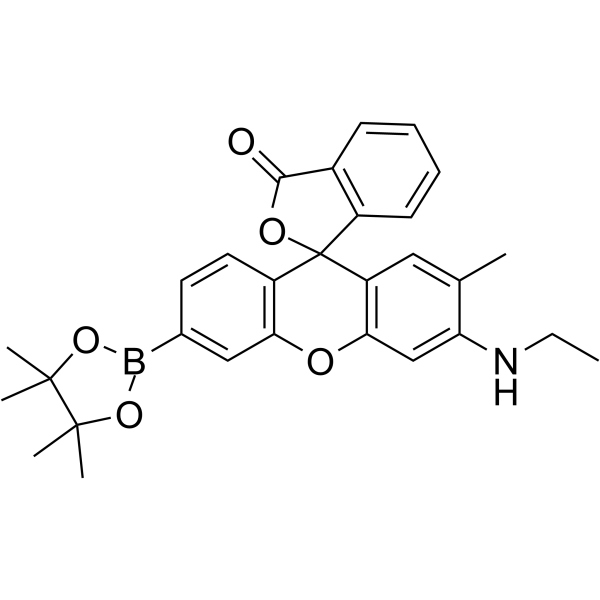
NucPE1;(Synonyms: Nuclear Peroxy Emerald 1) 纯度: ge;98.0%
NucPE1 (Nuclear Peroxy Emerald 1) 是核定位荧光过氧化氢探针, 其特异性定位于细胞核,没有附加的靶向部分。

NucPE1 Chemical Structure
CAS No. : 1404091-23-1
| 规格 | 价格 | 是否有货 | 数量 |
|---|---|---|---|
| 1 mg | ¥1500 | In-stock | |
| 5 mg | ¥4500 | In-stock | |
| 10 mg | ¥7500 | In-stock | |
| 50 mg | ¥21000 | In-stock | |
| 100 mg | ¥31000 | In-stock | |
| 200 mg | ; | 询价 | ; |
| 500 mg | ; | 询价 | ; |
* Please select Quantity before adding items.
NucPE1 相关产品
bull;相关化合物库:
- Bioactive Compound Library Plus
| 生物活性 |
NucPE1 (Nuclear Peroxy Emerald 1) is a nuclear-localized fluorescent hydrogen peroxide that is specifically localized to cellular nuclei without appended targeting moieties. |
||||||||||||||||
|---|---|---|---|---|---|---|---|---|---|---|---|---|---|---|---|---|---|
| 体外研究 (In Vitro) |
NucPE1 features two major visible region absorptions (λabs=468 nm, ε=27,300 M-1cm-1; λabs=490 nm, ε=26,000 M-1cm-1) and a weak emission (λem=530 nm, Φ=0.117). Reaction of NucPE1 with H2O2 triggers a fluorescence increase upon its conversion to fluorophore NucPE1, which possesses one major absorption band at 505 nm (ε=19,100 M-1cm-1) with enhanced emission (λem=530 nm, Φ=0.626). NucPE1 selectively accumulates in the nuclei of a variety of mammalian cell lines as well as in whole model organisms like C. clegans, where it can respond to subcellular changes in H2O2 fluxes[1]. MCE has not independently confirmed the accuracy of these methods. They are for reference only. |
||||||||||||||||
| 体内研究 (In Vivo) |
NucPE1 maintains the ability to selectively target nuclei in vivo. NucPE1 imaging reveals a reduction in nuclear H2O2 levels in worms overexpressing sir-2.1 compared to wildtype congeners, supporting a link between this longevity-promoting sirtuin protein and enhanced regulation of nuclear ROS pools[1]. MCE has not independently confirmed the accuracy of these methods. They are for reference only. |
||||||||||||||||
| 分子量 |
483.36 |
||||||||||||||||
| Formula |
C29H30BNO5 |
||||||||||||||||
| CAS 号 |
1404091-23-1 |
||||||||||||||||
| 运输条件 |
Room temperature in continental US; may vary elsewhere. |
||||||||||||||||
| 储存方式 |
4deg;C, protect from light, stored under nitrogen *In solvent : -80deg;C, 6 months; -20deg;C, 1 month (protect from light, stored under nitrogen) |
||||||||||||||||
| 溶解性数据 |
In Vitro:;
DMSO : 25 mg/mL (51.72 mM; Need ultrasonic) 配制储备液
*
请根据产品在不同溶剂中的溶解度选择合适的溶剂配制储备液;一旦配成溶液,请分装保存,避免反复冻融造成的产品失效。 In Vivo:
请根据您的实验动物和给药方式选择适当的溶解方案。以下溶解方案都请先按照 In Vitro 方式配制澄清的储备液,再依次添加助溶剂: ——为保证实验结果的可靠性,澄清的储备液可以根据储存条件,适当保存;体内实验的工作液,建议您现用现配,当天使用; 以下溶剂前显示的百
|
||||||||||||||||
| 参考文献 |
|
| Cell Assay [1][2] |
Excitation of NucPE1-loaded cells at 514 nm is carried out with an Ar laser and emission is collected using a META detector between 522–554 nm. Excitation of Hoechst 33342 is carried out using a MaiTai two-photon laser at 780-nm pulses and emission is collected between 436–501 nm. All images in an experiment are collected simultaneously using identical microscope settings. Image analysis is performed in Image J. The background fluorescence is measured in a data set by selecting an ROI outside of the cells. This background number is then used to set the threshold of the image and thereby select for NucPE1 signal. The mean fluorescence intensity of this signal is then measured and utilized as the fluorescent signal for that particular image. The same threshold settings are utilized for all images in an experiment[1]. The measurement of nuclear H2O2 is achieved using NucPE1. The cells are incubated for 45 min at 10 μM NucPE1 in the dark. The incubation is followed by ishing and analysis by flow cytometry as explained above. Mitochondrial ROS are measured using MitoSOX probe. The cells are incubated with 5 μM MitoSOX for 15 min in the dark. The incubation is followed by ishing and analysis by flow cytometry[2]. Live imaging of the NucPE1 probe is carried out using excitation at 488 nm with an argon laser, and emission is collected using a META detector at about 520 nm. The Hoechst dye is incubated together with NucPE1. When multiple staining of NucPE1 and PO1 is performed, the multitracking mode of scanning is applied for acquisition of the images[2]. MCE has not independently confirmed the accuracy of these methods. They are for reference only. |
|---|---|
| 参考文献 |
|
